| Product name | Smad4 Polyclonal Antibody |
| Immunogen | Synthesized peptide derived from the N-terminal region of human Smad4 at AA range: 40-120 |
| Host | Rabbit |
| Reactivity | Human,Mouse,Rat,Monkey |
| Applications | WB,IHC,IF,ELISA |
| Applications notes | Optimal working dilutions should be determined experimentally by the investigator. Suggested starting dilutions are as follows: WB 1:500-1:2000;IHC 1:100-1:300;IF 1:200-1:1000;ELISA 1:10000;Not yet tested in other applications. |
| Clonality | Polyclonal |
| Preparation method | The antibody was affinity-purified from rabbit antiserum by affinity-chromatography using epitope-specific immunogen. |
| Alternative | SMAD4; DPC4; MADH4; Mothers against decapentaplegic homolog 4; MAD homolog 4; Mothers against DPP homolog 4; Deletion target in pancreatic carcinoma 4; SMAD family member 4; SMAD 4; Smad4; hSMAD4 |
| Formulation | Liquid solution |
| Concentration | 1 mg/ml |
| Molecular weight | 60kD |
| Storage buffer | Liquid in PBS containing 50% glycerol, 0.5% BSA and 0.02% sodium azide. |
| Storage instructions | Stable for one year at -20°C from date of shipment. For maximum recovery of product, centrifuge the original vial after thawing and prior to removing the cap. Aliquot to avoid repeated freezing and thawing. |
| Shipping | Gel pack with blue ice. |
| Precautions | The product listed herein is for research use only and is not intended for use in human or clinical diagnosis. Suggested applications of our products are not recommendations to use our products in violation of any patent or as a license. We cannot be responsible for patent infringements or other violations that may occur with the use of this product. |
| Background | SMAD4 encodes a member of the Smad family of signal transduction proteins. Smad proteins are phosphorylated and activated by transmembrane serine-threonine receptor kinases in response to TGF-beta signaling. The product of SMAD4 forms homomeric complexes and heteromeric complexes with other activated Smad proteins, which then accumulate in the nucleus and regulate the transcription of target genes. This protein binds to DNA and recognizes an 8-bp palindromic sequence (GTCTAGAC) called the Smad-binding element (SBE). The Smad proteins are subject to complex regulation by post-translational modifications. Mutations or deletions in this gene have been shown to result in pancreatic cancer, juvenile polyposis syndrome, and hereditary hemorrhagic telangiectasia syndrome. |
| Gene ID | 4089 |
| Alternative | SMAD4; DPC4; MADH4; Mothers against decapentaplegic homolog 4; MAD homolog 4; Mothers against DPP homolog 4; Deletion target in pancreatic carcinoma 4; SMAD family member 4; SMAD 4; Smad4; hSMAD4 |
| Others | Smad4 Polyclonal Antibody detects endogenous levels of Smad4 protein. |
| Accession | Q13485 |
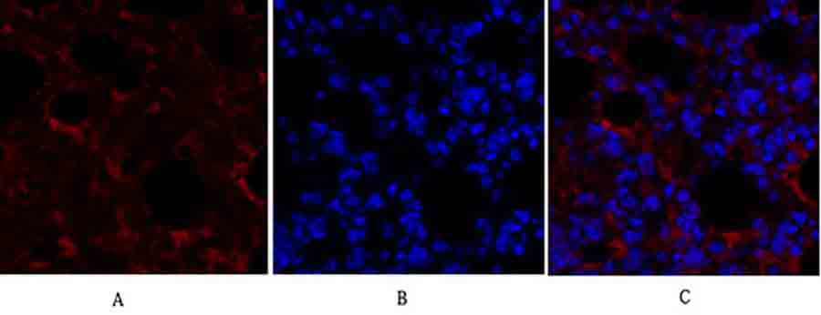
Fig.1. Immunofluorescence analysis of mouse lung tissue. 1, Smad4 Polyclonal Antibody (red) was diluted at 1:200 (4°C, overnight). 2, Cy3 Labeled secondary antibody was diluted at 1:300 (room temperature, 50min). 3, Picture B: DAPI (blue) 10min. Picture A: Target. Picture B: DAPI. Picture C: merge of A+B.
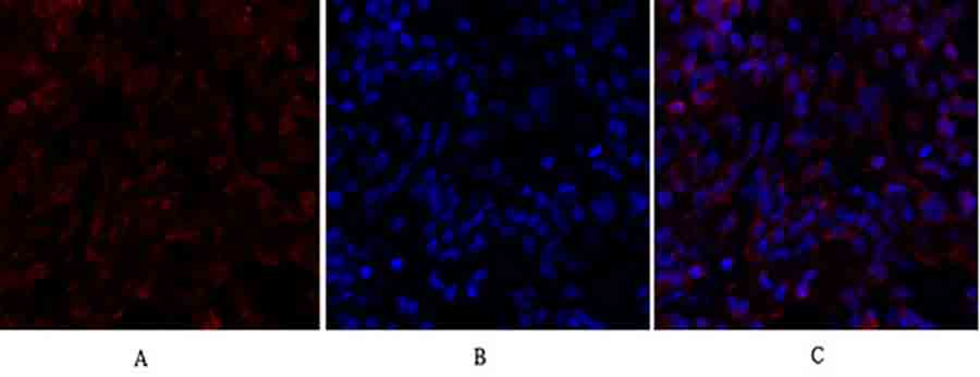
Fig.2. Immunofluorescence analysis of rat lung tissue. 1, Smad4 Polyclonal Antibody (red) was diluted at 1:200 (4°C, overnight). 2, Cy3 Labeled secondary antibody was diluted at 1:300 (room temperature, 50min). 3, Picture B: DAPI (blue) 10min. Picture A: Target. Picture B: DAPI. Picture C: merge of A+B.
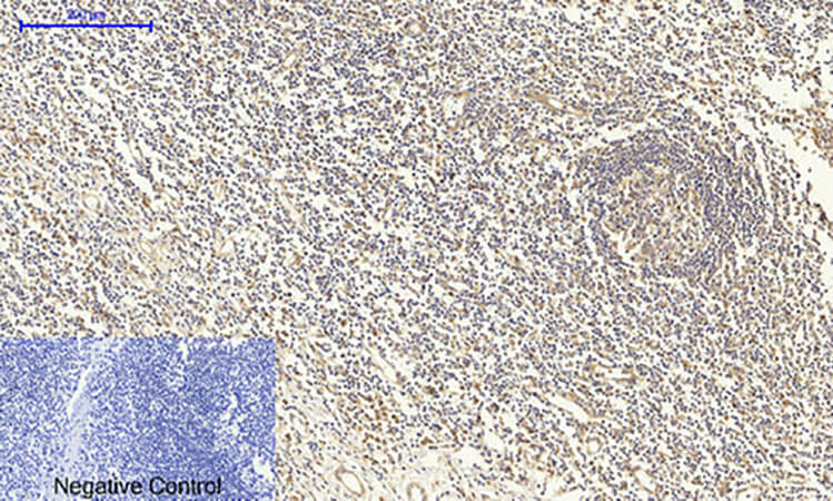
Fig.3. Immunohistochemical analysis of paraffin-embedded human tonsil tissue. 1, Smad4 Polyclonal Antibody was diluted at 1:200 (4°C, overnight). 2, Sodium citrate pH 6.0 was used for antibody retrieval (>98°C, 20min). 3, secondary antibody was diluted at 1:200 (room temperature, 30min). Negative control was used by secondary antibody only.
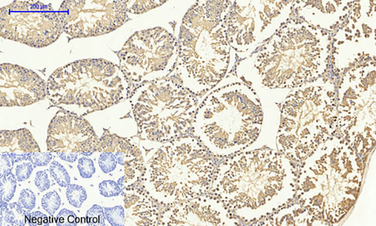
Fig.4. Immunohistochemical analysis of paraffin-embedded mouse testis tissue. 1, Smad4 Polyclonal Antibody was diluted at 1:200 (4°C, overnight). 2, Sodium citrate pH 6.0 was used for antibody retrieval (>98°C, 20min). 3, secondary antibody was diluted at 1:200 (room temperature, 30min). Negative control was used by secondary antibody only.
You must be logged in to post a review.


Reviews
There are no reviews yet.