| Product name | Smad2 Polyclonal Antibody |
| Immunogen | Synthesized peptide derived from human Smad2 around the non-phosphorylation site of S467 |
| Host | Rabbit |
| Reactivity | Human,Mouse,Rat |
| Applications | WB,CoIP,IHC,IF,ELISA |
| Applications notes | Optimal working dilutions should be determined experimentally by the investigator. Suggested starting dilutions are as follows: WB 1:500-1:2000;IHC 1:100-1:300;IF 1:200-1:1000;ELISA 1:5000;Not yet tested in other applications. |
| Clonality | Polyclonal |
| Preparation method | The antibody was affinity-purified from rabbit antiserum by affinity-chromatography using epitope-specific immunogen. |
| Alternative | SMAD2; MADH2; MADR2; Mothers against decapentaplegic homolog 2; MAD homolog 2; Mothers against DPP homolog 2; JV18-1; Mad-related protein 2; hMAD-2; SMAD family member 2; SMAD 2; Smad2; hSMAD2 |
| Formulation | Liquid solution |
| Concentration | 1 mg/ml |
| Molecular weight | 58kD |
| Storage buffer | Liquid in PBS containing 50% glycerol, 0.5% BSA and 0.02% sodium azide. |
| Storage instructions | Stable for one year at -20°C from date of shipment. For maximum recovery of product, centrifuge the original vial after thawing and prior to removing the cap. Aliquot to avoid repeated freezing and thawing. |
| Shipping | Gel pack with blue ice. |
| Precautions | The product listed herein is for research use only and is not intended for use in human or clinical diagnosis. Suggested applications of our products are not recommendations to use our products in violation of any patent or as a license. We cannot be responsible for patent infringements or other violations that may occur with the use of this product. |
| Background | The protein encoded by SMAD2 belongs to the SMAD, a family of proteins similar to the gene products of the Drosophila gene ‘mothers against decapentaplegic' (Mad) and the C. elegans gene Sma. SMAD proteins are signal transducers and transcriptional modulators that mediate multiple signaling pathways. This protein mediates the signal of the transforming growth factor (TGF)-beta, and thus regulates multiple cellular processes, such as cell proliferation, apoptosis, and differentiation. This protein is recruited to the TGF-beta receptors through its interaction with the SMAD anchor for receptor activation (SARA) protein. In response to TGF-beta signal, this protein is phosphorylated by the TGF-beta receptors. The phosphorylation induces the dissociation of this protein with SARA and the association with the family member SMAD4. The association with SMAD4 is important for the translocation of this protein into the nucleus, where it binds to target promoters and forms a transcription repressor complex with other cofactors. This protein can also be phosphorylated by activin type 1 receptor kinase, and mediates the signal from the activin. Alternatively spliced transcript variants have been observed for this gene. |
| Gene ID | 4087 |
| Alternative | SMAD2; MADH2; MADR2; Mothers against decapentaplegic homolog 2; MAD homolog 2; Mothers against DPP homolog 2; JV18-1; Mad-related protein 2; hMAD-2; SMAD family member 2; SMAD 2; Smad2; hSMAD2 |
| Others | Smad2 Polyclonal Antibody detects endogenous levels of Smad2 protein. |
| Accession | Q15796 |
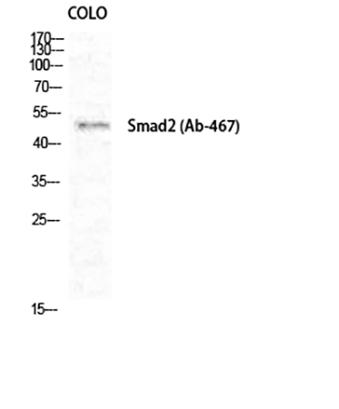
Fig.1. Western Blot analysis of COLO cells using Smad2 Polyclonal Antibody diluted at 1:1000.
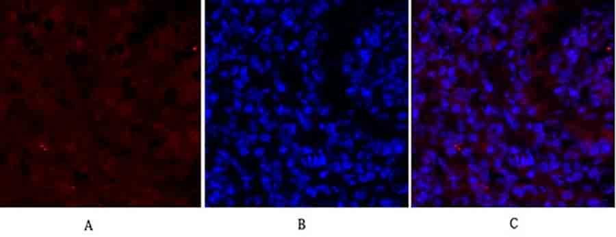
Fig.2. Immunofluorescence analysis of Mouse lung tissue. 1, Smad2 Polyclonal Antibody (red) was diluted at 1:200 (4°C, overnight). 2, Cy3 labled secondary antibody was diluted at 1:300 (room temperature, 50min). 3, Picture B: DAPI (blue) 10min. Picture A: Target. Picture B: DAPI. Picture C: merge of A+B.
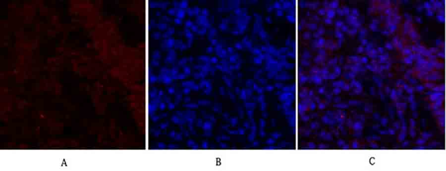
Fig.3. Immunofluorescence analysis of rat lung tissue. 1, Smad2 Polyclonal Antibody (red) was diluted at 1:200 (4°C, overnight). 2, Cy3 labled secondary antibody was diluted at 1:300 (room temperature, 50min). 3, Picture B: DAPI (blue) 10min. Picture A: Target. Picture B: DAPI. Picture C: merge of A+B.
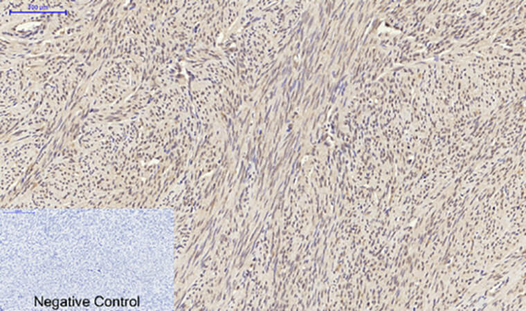
Fig.4. Immunohistochemical analysis of paraffin-embedded human uterus tissue. 1, Smad2 Polyclonal Antibody was diluted at 1:200 (4°C, overnight). 2, Sodium citrate pH 6.0 was used for antibody retrieval (>98°C, 20min). 3, secondary antibody was diluted at 1:200 (room temperature, 30min). Negative control was used by secondary antibody only.
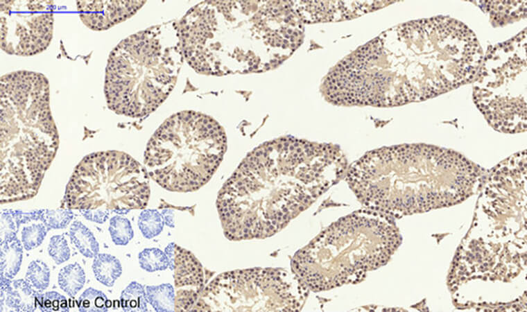
Fig.5. Immunohistochemical analysis of paraffin-embedded Mouse testis tissue. 1, Smad2 Polyclonal Antibody was diluted at 1:200 (4°C, overnight). 2, Sodium citrate pH 6.0 was used for antibody retrieval (>98°C, 20min). 3, secondary antibody was diluted at 1:200 (room temperature, 30min). Negative control was used by secondary antibody only.
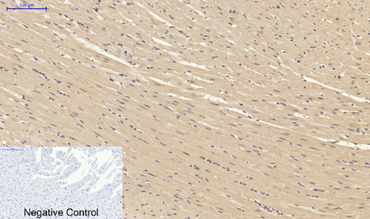
Fig.6. Immunohistochemical analysis of paraffin-embedded rat heart tissue. 1, Smad2 Polyclonal Antibody was diluted at 1:200 (4°C, overnight). 2, Sodium citrate pH 6.0 was used for antibody retrieval (>98°C, 20min). 3, secondary antibody was diluted at 1:200 (room temperature, 30min). Negative control was used by secondary antibody only.
You must be logged in to post a review.


Reviews
There are no reviews yet.