| Product name | SCCA1/2 Polyclonal Antibody |
| Immunogen | Synthesized peptide derived from the Internal region of human SCCA1/2 |
| Host | Rabbit |
| Reactivity | Human |
| Applications | WB,IHC,IF,ELISA |
| Applications notes | Optimal working dilutions should be determined experimentally by the investigator. Suggested starting dilutions are as follows: WB 1:500-1:2000;IHC: 1:100-1:300;ELISA 1:20000;IF 1:50-200 |
| Clonality | Polyclonal |
| Preparation method | The antibody was affinity-purified from rabbit antiserum by affinity-chromatography using epitope-specific immunogen. |
| Alternative | SERPINB3; SCCA; SCCA1; Serpin B3; Protein T4-A; Squamous cell carcinoma antigen 1; SCCA-1 |
| Formulation | Liquid solution |
| Concentration | 1 mg/ml |
| Molecular weight | 45kD |
| Storage buffer | Liquid in PBS containing 50% glycerol, 0.5% BSA and 0.02% sodium azide. |
| Storage instructions | Stable for one year at -20°C from date of shipment. For maximum recovery of product, centrifuge the original vial after thawing and prior to removing the cap. Aliquot to avoid repeated freezing and thawing. |
| Shipping | Gel pack with blue ice. |
| Precautions | The product listed herein is for research use only and is not intended for use in human or clinical diagnosis. Suggested applications of our products are not recommendations to use our products in violation of any patent or as a license. We cannot be responsible for patent infringements or other violations that may occur with the use of this product. |
| Background | May act as a protease inhibitor to modulate the host immune response against tumor cells. |
| Gene ID | 6317 |
| Alternative | SERPINB3; SCCA; SCCA1; Serpin B3; Protein T4-A; Squamous cell carcinoma antigen 1; SCCA-1 |
| Others | SCCA1/2 Polyclonal Antibody detects endogenous levels of SCCA1/2 protein. |
| Accession | P29508 |
| Observed Band(KD) | 45 |
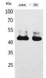
Fig.1. Western Blot analysis of Jurkat, 293 cells using SCCA1/2 Polyclonal Antibody. Secondary antibody (catalog#: A21020) was diluted at 1:20000.
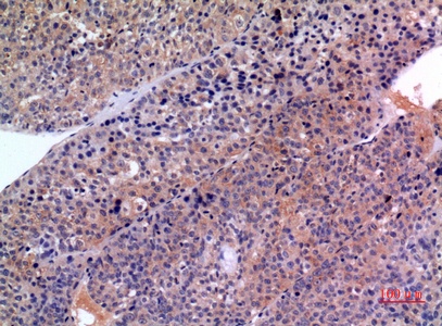
Fig.2. Immunohistochemical analysis of paraffin-embedded human-liver-cancer, antibody was diluted at 1:100.
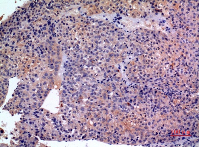
Fig.3. Immunohistochemical analysis of paraffin-embedded human-liver-cancer, antibody was diluted at 1:100.
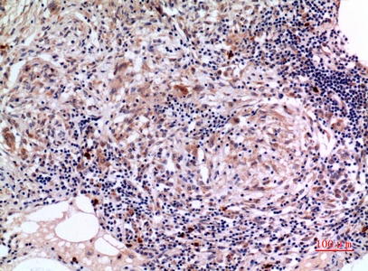
Fig.4. Immunohistochemical analysis of paraffin-embedded human-lung, antibody was diluted at 1:100.

Fig.5. Immunohistochemical analysis of paraffin-embedded human-lung, antibody was diluted at 1:100.
You must be logged in to post a review.


Reviews
There are no reviews yet.