| Product name | PPAR-γ Polyclonal Antibody |
| Immunogen | Synthesized peptide derived from human PPAR-γ around the non-phosphorylation site of S112 |
| Host | Rabbit |
| Reactivity | Human, Mouse, Rat |
| Applications | ELISA, IF, IHC-P, WB |
| Applications notes | Optimal working dilutions should be determined experimentally by the investigator. Suggested starting dilutions are as follows: WB (1:500-1:2000), IHC-P (1:100-1:300), ELISA (1:10000). Not yet tested in other applications. |
| Clonality | Polyclonal |
| Preparation method | The antibody was affinity-purified from rabbit antiserum by affinity-chromatography using epitope-specific immunogen |
| Alternative | PPARG; NR1C3; Peroxisome proliferator-activated receptor gamma; PPAR-gamma; Nuclear receptor subfamily 1 group C member 3 |
| Formulation | Liquid solution |
| Concentration | 1 mg/ml |
| Storage buffer | PBS containing 50% Glycerol, 0.5% BSA and 0.02% Sodium Azide. |
| Storage instructions | Stable for one year at -20°C from date of shipment. For maximum recovery of product, centrifuge the original vial after thawing and prior to removing the cap. Aliquot to avoid repeated freezing and thawing. |
| Shipping | Gel pack with blue ice. |
| Precautions | The product listed herein is for research use only and is not intended for use in human or clinical diagnosis. Suggested applications of our products are not recommendations to use our products in violation of any patent or as a license. We cannot be responsible for patent infringements or other violations that may occur with the use of this product. |
| Background | PPARG encodes a member of the peroxisome proliferator-activated receptor (PPAR) subfamily of nuclear receptors. PPARs form heterodimers with retinoid X receptors (RXRs) and these heterodimers regulate transcription of various genes. Three subtypes of PPARs are known: PPAR-alpha, PPAR-delta, and PPAR-gamma. Peroxisome proliferator activated receptor gamma encoded by PPARG is PPAR-gamma and is a regulator of adipocyte differentiation. Additionally, PPAR-gamma has been implicated in the pathology of numerous diseases including obesity, diabetes, atherosclerosis and cancer. Alternatively spliced transcript variants that encode different isoforms have been described |
| Gene ID | 5468 |
| Alternative | PPARG; NR1C3; Peroxisome proliferator-activated receptor gamma; PPAR-gamma; Nuclear receptor subfamily 1 group C member 3 |
| Others | PPAR-γ Polyclonal Antibody detects endogenous levels of PPAR-γ protein. |
| Accession | P37231 |
| Observed Band(KD) | 57 |
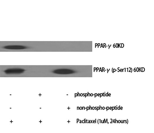
Fig.1. Western Blot analysis of various cells using PPAR-γ Polyclonal Antibody diluted at 1:1000.
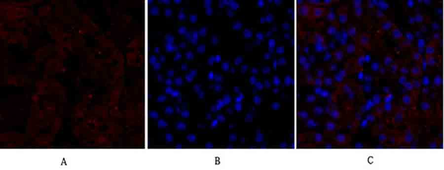
Fig.2. Immunofluorescence analysis of mouse kidney tissue. 1, PPAR-γ Polyclonal Antibody (red) was diluted at 1:200 (4°C, overnight). 2, Cy3 Labeled secondary antibody was diluted at 1:300 (room temperature, 50min). 3, Picture B: DAPI (blue) 10min. Picture A: Target. Picture B: DAPI. Picture C: merge of A+B.
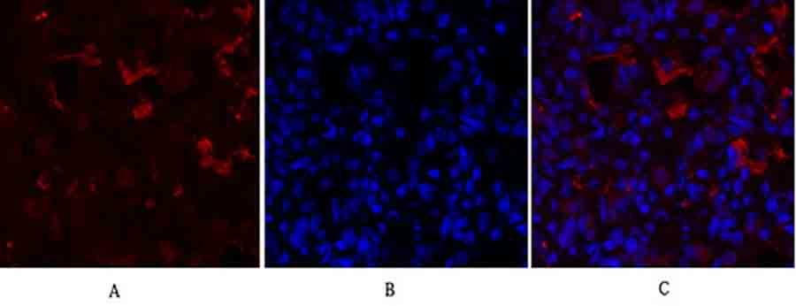
Fig.3. Immunofluorescence analysis of rat lung tissue. 1, PPAR-γ Polyclonal Antibody (red) was diluted at 1:200 (4°C, overnight). 2, Cy3 Labeled secondary antibody was diluted at 1:300 (room temperature, 50min). 3, Picture B: DAPI (blue) 10min. Picture A: Target. Picture B: DAPI. Picture C: merge of A+B.
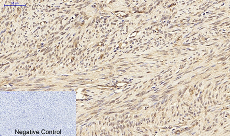
Fig.4. Immunohistochemical analysis of paraffin-embedded human uterus tissue. 1, PPAR-γ Polyclonal Antibody was diluted at 1:200 (4°C, overnight). 2, Sodium citrate pH 6.0 was used for antibody retrieval (>98°C, 20min). 3, secondary antibody was diluted at 1:200 (room temperature, 30min). Negative control was used by secondary antibody only.
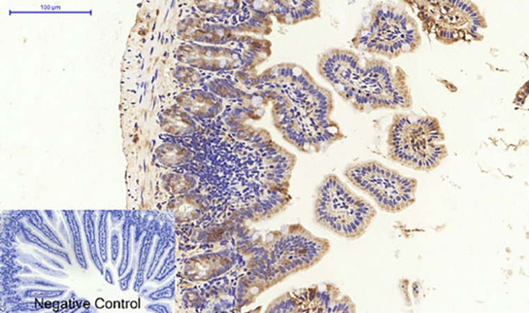
Fig.5. Immunohistochemical analysis of paraffin-embedded mouse colon tissue. 1, PPAR-γ Polyclonal Antibody was diluted at 1:200 (4°C, overnight). 2, Sodium citrate pH 6.0 was used for antibody retrieval (>98°C, 20min). 3, secondary antibody was diluted at 1:200 (room temperature, 30min). Negative control was used by secondary antibody only.
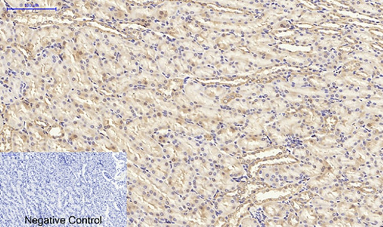
Fig.6. Immunohistochemical analysis of paraffin-embedded rat kidney tissue. 1, PPAR-γ Polyclonal Antibody was diluted at 1:200 (4°C, overnight). 2, Sodium citrate pH 6.0 was used for antibody retrieval (>98°C, 20min). 3, secondary antibody was diluted at 1:200 (room temperature, 30min). Negative control was used by secondary antibody only.
Author:BK Mishra, BD Banerjee, V Agrawal Publication name:Endocrine IF:2.55
Author:Hatiboglu S, Yanar F, Ozturk A, et al Publication name:Turkish Journal of Biochemistry IF:0.7
You must be logged in to post a review.
1.The species of antibody reactivity should be the sample species that can be matched normally after Abbkine R&D experts have passed strict scientific verification. If your sample is not within the range of reactivity, in order to improve the efficiency and results of your experiment, it is not suggested to try other species. Otherwise, it may lead to sample mismatch and affect the effect of your experiment.
2.Please aliquot the antibody received as soon as possible and store it at -20℃, avoid repeated freezing and thawing, and use it within one year.
Welcome any form of communications, and better service will be provided here.
Tell: +1-404-854-0155
Email: service@abbkine.com
Support Email: support@abbkine.com
Address: 3052 Stroop Hill Road, Apt 203, Atlanta 30303, Georgia, United States of America


Reviews
There are no reviews yet.