| Product name | PPAR-α Polyclonal Antibody |
| Immunogen | Synthesized peptide derived from human PPAR-α around the non-phosphorylation site of S21 |
| Host | Rabbit |
| Reactivity | Human,Mouse,Rat |
| Applications | WB,IHC,IF,ELISA |
| Applications notes | Optimal working dilutions should be determined experimentally by the investigator. Suggested starting dilutions are as follows: WB 1:500-2000;ELISA 1:10000-20000;IHC 1:50-300;IF 1:50-200 |
| Clonality | Polyclonal |
| Preparation method | The antibody was affinity-purified from rabbit antiserum by affinity-chromatography using epitope-specific immunogen. |
| Alternative | PPARA; NR1C1; PPAR; Peroxisome proliferator-activated receptor alpha; PPAR-alpha; Nuclear receptor subfamily 1 group C member 1 |
| Formulation | Liquid solution |
| Concentration | 1 mg/ml |
| Molecular weight | 52kD |
| Storage buffer | Liquid in PBS containing 50% glycerol, 0.5% BSA and 0.02% sodium azide. |
| Storage instructions | Stable for one year at -20°C from date of shipment. For maximum recovery of product, centrifuge the original vial after thawing and prior to removing the cap. Aliquot to avoid repeated freezing and thawing. |
| Shipping | Gel pack with blue ice. |
| Precautions | The product listed herein is for research use only and is not intended for use in human or clinical diagnosis. Suggested applications of our products are not recommendations to use our products in violation of any patent or as a license. We cannot be responsible for patent infringements or other violations that may occur with the use of this product. |
| Background | Peroxisome proliferators include hypolipidemic drugs, herbicides, leukotriene antagonists, and plasticizers; this term arises because they induce an increase in the size and number of peroxisomes. Peroxisomes are subcellular organelles found in plants and animals that contain enzymes for respiration and for cholesterol and lipid metabolism. The action of peroxisome proliferators is thought to be mediated via specific receptors, called PPARs, which belong to the steroid hormone receptor superfamily. PPARs affect the expression of target genes involved in cell proliferation, cell differentiation and in immune and inflammation responses. Three closely related subtypes (alpha, beta/delta, and gamma) have been identified. PPARA (peroxisome proliferator activated receptor alpha) encodes the subtype PPAR-alpha, which is a nuclear transcription factor. Multiple alternatively spliced transcript variants have been described for PPARA, although the full-length nature of only two has been determined. |
| Gene ID | 5465 |
| Alternative | PPARA; NR1C1; PPAR; Peroxisome proliferator-activated receptor alpha; PPAR-alpha; Nuclear receptor subfamily 1 group C member 1 |
| Others | PPAR-α Polyclonal Antibody detects endogenous levels of PPAR-α protein. |
| Accession | Q07869 |
| Observed Band(KD) | 52 |
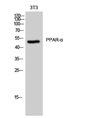
Fig.1. Western Blot analysis of 3T3 cells using PPAR-α Polyclonal Antibody.
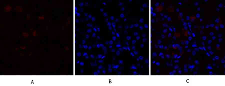
Fig.2. Immunofluorescence analysis of mouse kidney tissue. 1, PPAR-α Polyclonal Antibody (red) was diluted at 1:200 (4°C, overnight). 2, Cy3 Labeled secondary antibody was diluted at 1:300 (room temperature, 50min). 3, Picture B: DAPI (blue) 10min. Picture A: Target. Picture B: DAPI. Picture C: merge of A+B.
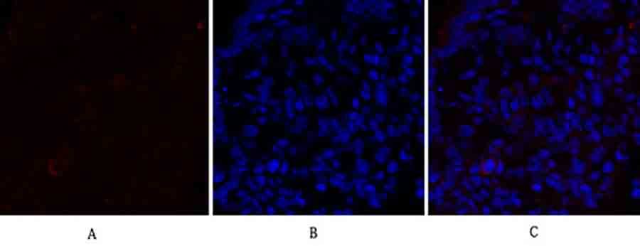
Fig.3. Immunofluorescence analysis of rat lung tissue. 1, PPAR-α Polyclonal Antibody (red) was diluted at 1:200 (4°C, overnight). 2, Cy3 Labeled secondary antibody was diluted at 1:300 (room temperature, 50min). 3, Picture B: DAPI (blue) 10min. Picture A: Target. Picture B: DAPI. Picture C: merge of A+B.
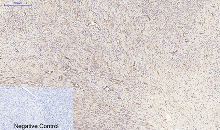
Fig.4. Immunohistochemical analysis of paraffin-embedded human uterus cancer tissue. 1, PPAR-α Polyclonal Antibody was diluted at 1:200 (4°C, overnight). 2, Sodium citrate pH 6.0 was used for antibody retrieval (>98°C, 20min). 3, secondary antibody was diluted at 1:200 (room temperature, 30min). Negative control was used by secondary antibody only.
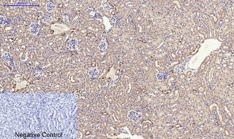
Fig.5. Immunohistochemical analysis of paraffin-embedded mouse kidney tissue. 1, PPAR-α Polyclonal Antibody was diluted at 1:200 (4°C, overnight). 2, Sodium citrate pH 6.0 was used for antibody retrieval (>98°C, 20min). 3, secondary antibody was diluted at 1:200 (room temperature, 30min). Negative control was used by secondary antibody only.
Author:Mi A, Hu Q, Liu Y Publication name:Food & Function IF:6
Author:Zhang Y, Li T Publication name:J Transl Med IF:4
You must be logged in to post a review.


Reviews
There are no reviews yet.