| Product name | PERK Polyclonal Antibody |
| Immunogen | Synthesized peptide derived from human PERK around the non-phosphorylation site of T981 |
| Host | Rabbit |
| Reactivity | Human,Mouse,Rat |
| Applications | IF,WB,IHC,ELISA |
| Applications notes | Optimal working dilutions should be determined experimentally by the investigator. Suggested starting dilutions are as follows: IF 1:50-200;WB 1:500-1:2000;IHC 1:100-1:300;ELISA 1:40000;Not yet tested in other applications; |
| Clonality | Polyclonal |
| Preparation method | The antibody was affinity-purified from rabbit antiserum by affinity-chromatography using epitope-specific immunogen. |
| Alternative | EIF2AK3; PEK; PERK; Eukaryotic translation initiation factor 2-alpha kinase 3; PRKR-like endoplasmic reticulum kinase; Pancreatic eIF2-alpha kinase; HsPEK |
| Formulation | Liquid solution |
| Concentration | 1 mg/ml |
| Molecular weight | 125kD |
| Storage buffer | Liquid in PBS containing 50% glycerol, 0.5% BSA and 0.02% sodium azide. |
| Storage instructions | Stable for one year at -20°C from date of shipment. For maximum recovery of product, centrifuge the original vial after thawing and prior to removing the cap. Aliquot to avoid repeated freezing and thawing. |
| Shipping | Gel pack with blue ice. |
| Precautions | The product listed herein is for research use only and is not intended for use in human or clinical diagnosis. Suggested applications of our products are not recommendations to use our products in violation of any patent or as a license. We cannot be responsible for patent infringements or other violations that may occur with the use of this product. |
| Background | The protein encoded by EIF2AK3 (eukaryotic translation initiation factor 2 alpha kinase 3) phosphorylates the alpha subunit of eukaryotic translation-initiation factor 2, leading to its inactivation, and thus to a rapid reduction of translational initiation and repression of global protein synthesis. This protein is thought to modulate mitochondrial function. It is a type I membrane protein located in the endoplasmic reticulum (ER), where it is induced by ER stress caused by malfolded proteins. Mutations in EIF2AK3 are associated with Wolcott-Rallison syndrome. |
| Gene ID | 9451 |
| Alternative | EIF2AK3; PEK; PERK; Eukaryotic translation initiation factor 2-alpha kinase 3; PRKR-like endoplasmic reticulum kinase; Pancreatic eIF2-alpha kinase; HsPEK |
| Others | PERK Polyclonal Antibody detects endogenous levels of PERK protein. |
| Accession | Q9NZJ5 |
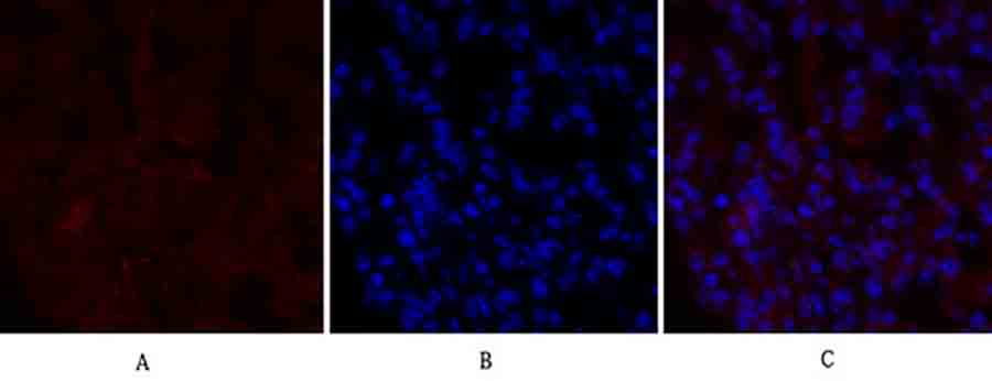
Fig.1. Immunofluorescence analysis of mouse lung tissue. 1, PERK Polyclonal Antibody (red) was diluted at 1:200 (4°C, overnight). 2, Cy3 Labeled secondary antibody was diluted at 1:300 (room temperature, 50min). 3, Picture B: DAPI (blue) 10min. Picture A: Target. Picture B: DAPI. Picture C: merge of A+B.
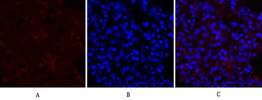
Fig.2. Immunofluorescence analysis of rat lung tissue. 1, PERK Polyclonal Antibody (red) was diluted at 1:200 (4°C, overnight). 2, Cy3 Labeled secondary antibody was diluted at 1:300 (room temperature, 50min). 3, Picture B: DAPI (blue) 10min. Picture A: Target. Picture B: DAPI. Picture C: merge of A+B.
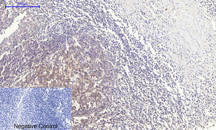
Fig.3. Immunohistochemical analysis of paraffin-embedded human tonsil tissue. 1, PERK Polyclonal Antibody was diluted at 1:200 (4°C, overnight). 2, Sodium citrate pH 6.0 was used for antibody retrieval (>98°C, 20min). 3, secondary antibody was diluted at 1:200 (room temperature, 30min). Negative control was used by secondary antibody only.
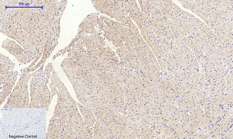
Fig.4. Immunohistochemical analysis of paraffin-embedded mouse heart tissue. 1, PERK Polyclonal Antibody was diluted at 1:200 (4°C, overnight). 2, Sodium citrate pH 6.0 was used for antibody retrieval (>98°C, 20min). 3, secondary antibody was diluted at 1:200 (room temperature, 30min). Negative control was used by secondary antibody only.
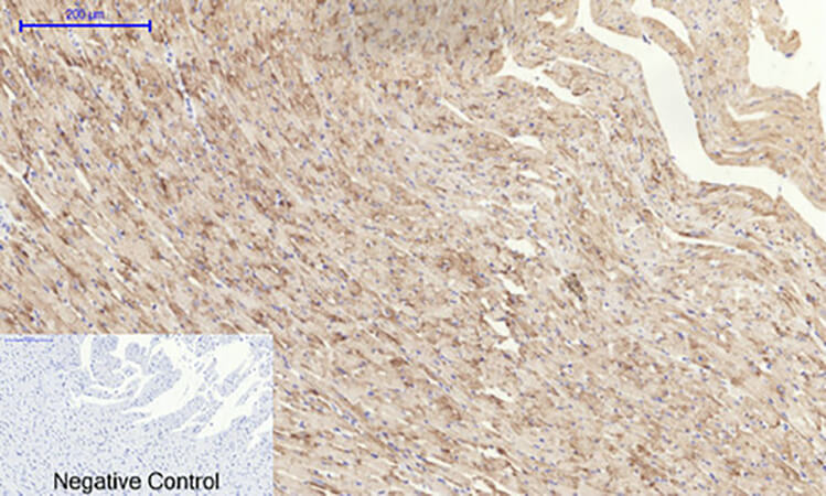
Fig.5. Immunohistochemical analysis of paraffin-embedded rat heart tissue. 1, PERK Polyclonal Antibody was diluted at 1:200 (4°C, overnight). 2, Sodium citrate pH 6.0 was used for antibody retrieval (>98°C, 20min). 3, secondary antibody was diluted at 1:200 (room temperature, 30min). Negative control was used by secondary antibody only.
Author:Liu W, Wang L, Zhang J, Qiao L, Liu Y, Yang X, Zhang J, Zheng W, Ma Z Publication name:3 Biotech IF:2
You must be logged in to post a review.


Reviews
There are no reviews yet.