| Product name | OPG Polyclonal Antibody |
| Immunogen | Synthesized peptide derived from the N-terminal region of human OPG at AA range: 10-90 |
| Host | Rabbit |
| Reactivity | Human,Mouse |
| Applications | WB,IHC,IF,ELISA |
| Applications notes | Optimal working dilutions should be determined experimentally by the investigator. Suggested starting dilutions are as follows: WB 1:500-1:2000;IHC: 1:100-300;ELISA 1:20000;IF 1:100-300;Not yet tested in other applications. |
| Clonality | Polyclonal |
| Preparation method | The antibody was affinity-purified from rabbit antiserum by affinity-chromatography using epitope-specific immunogen. |
| Alternative | TNFRSF11B; OCIF; OPG; Tumor necrosis factor receptor superfamily member 11B; Osteoclastogenesis inhibitory factor; Osteoprotegerin |
| Formulation | Liquid solution |
| Concentration | 1 mg/ml |
| Molecular weight | 55kD |
| Storage buffer | Liquid in PBS containing 50% glycerol, 0.5% BSA and 0.02% sodium azide. |
| Storage instructions | Stable for one year at -20°C from date of shipment. For maximum recovery of product, centrifuge the original vial after thawing and prior to removing the cap. Aliquot to avoid repeated freezing and thawing. |
| Shipping | Gel pack with blue ice. |
| Precautions | The product listed herein is for research use only and is not intended for use in human or clinical diagnosis. Suggested applications of our products are not recommendations to use our products in violation of any patent or as a license. We cannot be responsible for patent infringements or other violations that may occur with the use of this product. |
| Background | TNF receptor superfamily member 11b encoded by TNFRSF11B is a member of the TNF-receptor superfamily. TNF receptor superfamily member 11b is an osteoblast-secreted decoy receptor that functions as a negative regulator of bone resorption. TNF receptor superfamily member 11b specifically binds to its ligand, osteoprotegerin ligand, both of which are key extracellular regulators of osteoclast development. Studies of the mouse counterpart also suggest that TNF receptor superfamily member 11b and its ligand play a role in lymph-node organogenesis and vascular calcification. Alternatively spliced transcript variants of TNFRSF11B have been reported, but their full length nature has not been determined. |
| Gene ID | 4982 |
| Alternative | TNFRSF11B; OCIF; OPG; Tumor necrosis factor receptor superfamily member 11B; Osteoclastogenesis inhibitory factor; Osteoprotegerin |
| Others | OPG Polyclonal Antibody detects endogenous levels of OPG protein. |
| Accession | O00300 |
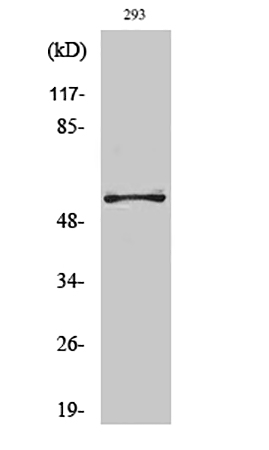
Fig.1. Western Blot analysis of various cells using OPG Polyclonal Antibody diluted at 1:1000.
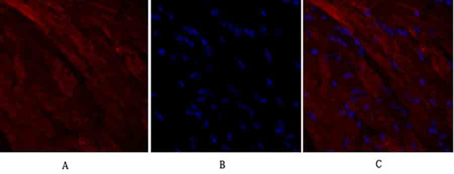
Fig.2. Immunofluorescence analysis of mouse heart tissue. 1, OPG Polyclonal Antibody (red) was diluted at 1:200 (4°C, overnight). 2, Cy3 Labeled secondary antibody was diluted at 1:300 (room temperature, 50min). 3, Picture B: DAPI (blue) 10min. Picture A: Target. Picture B: DAPI. Picture C: merge of A+B.
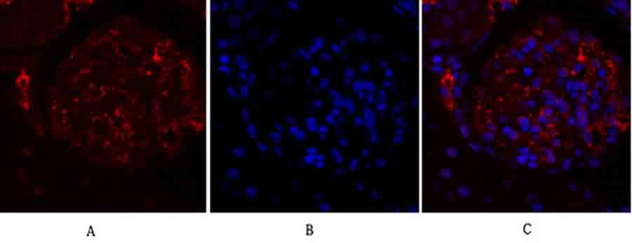
Fig.3. Immunofluorescence analysis of rat kidney tissue. 1, OPG Polyclonal Antibody (red) was diluted at 1:200 (4°C, overnight). 2, Cy3 Labeled secondary antibody was diluted at 1:300 (room temperature, 50min). 3, Picture B: DAPI (blue) 10min. Picture A: Target. Picture B: DAPI. Picture C: merge of A+B.
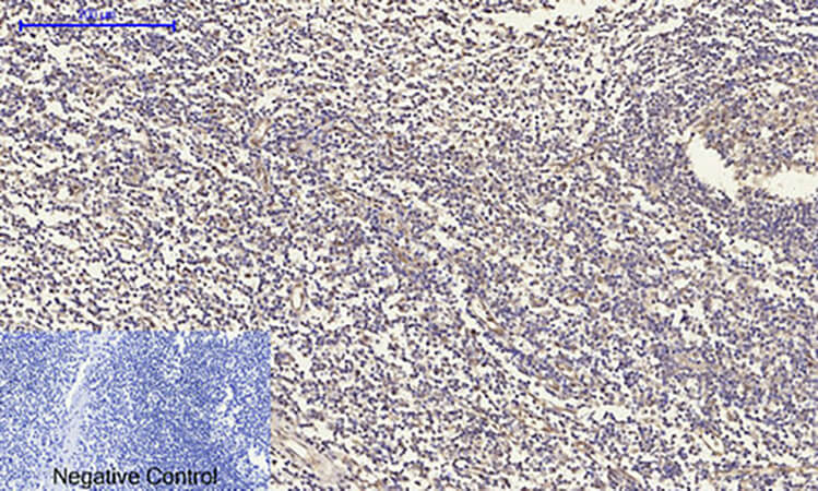
Fig.4. Immunohistochemical analysis of paraffin-embedded human tonsil tissue. 1, OPG Polyclonal Antibody was diluted at 1:200 (4°C, overnight). 2, Sodium citrate pH 6.0 was used for antibody retrieval (>98°C, 20min). 3, secondary antibody was diluted at 1:200 (room temperature, 30min). Negative control was used by secondary antibody only.
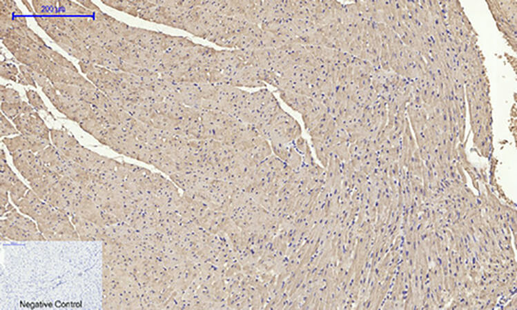
Fig.5. Immunohistochemical analysis of paraffin-embedded mouse heart tissue. 1, OPG Polyclonal Antibody was diluted at 1:200 (4°C, overnight). 2, Sodium citrate pH 6.0 was used for antibody retrieval (>98°C, 20min). 3, secondary antibody was diluted at 1:200 (room temperature, 30min). Negative control was used by secondary antibody only.
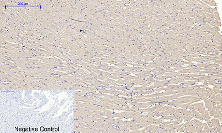
Fig.6. Immunohistochemical analysis of paraffin-embedded rat heart tissue. 1, OPG Polyclonal Antibody was diluted at 1:200 (4°C, overnight). 2, Sodium citrate pH 6.0 was used for antibody retrieval (>98°C, 20min). 3, secondary antibody was diluted at 1:200 (room temperature, 30min). Negative control was used by secondary antibody only.
You must be logged in to post a review.


Reviews
There are no reviews yet.