| Product name | Oct1 Monoclonal Antibody |
| Immunogen | Synthetic Peptide |
| Host | Mouse |
| Reactivity | Human |
| Applications | WB,IHC,IF, |
| Applications notes | Optimal working dilutions should be determined experimentally by the investigator. Suggested starting dilutions are as follows: WB 1:500-2000;IF 1:200;IHC 1:50-300 |
| Clonality | Monoclonal |
| Preparation method | The antibody was affinity-purified from mouse ascites by affinity-chromatography using specific immunogen. |
| Formulation | Liquid solution |
| Concentration | 1 mg/ml |
| Molecular weight | 89kD |
| Storage buffer | PBS, pH 7.4, containing 0.5%BSA, 0.02% sodium azide as Preservative and 50% Glycerol. |
| Storage instructions | Stable for one year at -20°C from date of shipment. For maximum recovery of product, centrifuge the original vial after thawing and prior to removing the cap. Aliquot to avoid repeated freezing and thawing. |
| Shipping | Gel pack with blue ice. |
| Precautions | The product listed herein is for research use only and is not intended for use in human or clinical diagnosis. Suggested applications of our products are not recommendations to use our products in violation of any patent or as a license. We cannot be responsible for patent infringements or other violations that may occur with the use of this product. |
| Background | The OCT1 transcription factor was among the first identified members of the POU transcription factor family (summarized by Sturm et al., 1993 [PubMed 8314572]). Members of this family contain the POU domain, a 160-amino acid region necessary for DNA binding to the octameric sequence ATGCAAAT. |
| Gene ID | 5451/5452 |
| Others | The antibody detects endogenous Oct1 protein. |
| Accession | P14859/P09086 |
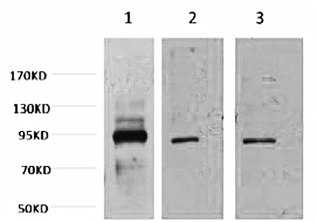
Fig.1. Western blot analysis of 1) Hela, 2) Jurkat, 3) HepG2, diluted at 1:2000
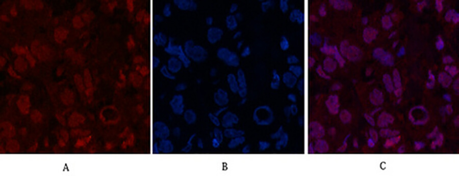
Fig.2. Immunofluorescence analysis of human breast cancer tissue. 1, Oct1 Monoclonal Antibody (red) was diluted at 1:200 (4°C, overnight). 2, Cy3 Labeled secondary antibody was diluted at 1:300 (room temperature, 50min). 3, Picture B: DAPI (blue) 10min. Picture A: Target. Picture B: DAPI. Picture C: merge of A+B.
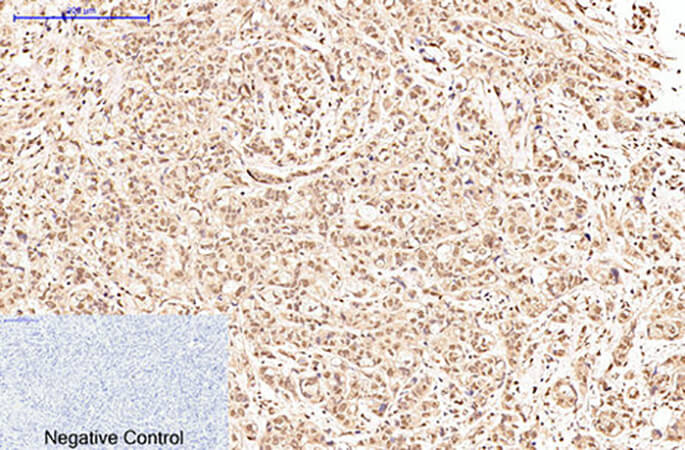
Fig.3. Immunohistochemical analysis of paraffin-embedded human breast cancer tissue. 1, Oct1 Monoclonal Antibody was diluted at 1:200 (4°C, overnight). 2, Sodium citrate pH 6.0 was used for antibody retrieval (>98°C, 20min). 3, secondary antibody was diluted at 1:200 (room temperature, 30min). Negative control was used by secondary antibody only.
You must be logged in to post a review.


Reviews
There are no reviews yet.