| Product name | MICU1 Monoclonal Antibody |
| Immunogen | Recombinant Protein |
| Host | Mouse |
| Reactivity | Human,Mouse,Rat |
| Applications | WB,IF,IHC |
| Applications notes | Optimal working dilutions should be determined experimentally by the investigator. Suggested starting dilutions are as follows: WB 1:1000-2000;IHC 1:100-200;IF 1:200 |
| Clonality | Monoclonal |
| Preparation method | The antibody was affinity-purified from mouse ascites by affinity-chromatography using epitope-specific immunogen. |
| Formulation | Liquid solution |
| Concentration | 1 mg/ml |
| Molecular weight | 55kD |
| Storage buffer | PBS, pH 7.4, containing 0.5%BSA, 0.02% sodium azide as Preservative and 50% Glycerol. |
| Storage instructions | Stable for one year at -20°C from date of shipment. For maximum recovery of product, centrifuge the original vial after thawing and prior to removing the cap. Aliquot to avoid repeated freezing and thawing. |
| Shipping | Gel pack with blue ice. |
| Precautions | The product listed herein is for research use only and is not intended for use in human or clinical diagnosis. Suggested applications of our products are not recommendations to use our products in violation of any patent or as a license. We cannot be responsible for patent infringements or other violations that may occur with the use of this product. |
| Background | Most PKMT substrates are histone proteins and transcription factors, emphasizing the importance of lysine methylation in regulating chromatin structure and gene expression. Lys9 of histone H3 is mono- or di-methylated by G9A/GLP and tri-methylated by SETDB1 to activate transcription. JHDM3A-mediated demethylation of the same residue creates mono-methyl Lys9 and inhibits gene transcription . Tumor suppressor p53 is regulated by methylation of at least four sites. p53-mediated transcription is repressed following mono-methylation of p53 at Lys370 by SMYD2; di-methylation at the same residue further inhibits p53 by preventing association with 53BP1. Concomitant di-methylation at Lys382 inhibits p53 ubiquitination following DNA damage. Mono-methylation at Lys382 by SET8 suppresses p53 transcriptional activity, while SET7/9 mono-methylation at Lys372 inhibits SMYD2 methylation at Lys370 and stabilizes the p53 protein. Di-methylation at Lys373 by G9A/GLP inhibits p53-mediated apoptosis and correlates with tri-methylation of histone H3 Lys9 at the p21 promoter . Overexpression of PKMTs is associated with multiple forms of human cancer, which has generated tremendous interest in targeting protein lysine methyltransferases in drug discovery research. |
| Gene ID | 10367 |
| Others | The antibody detects endogenous MICU1 protein. |
| Accession | Q9BPX6 |
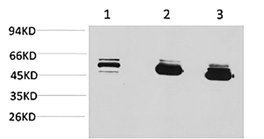
Fig.1. Western blot analysis of 1) MCF7, 2) mouse brain tissue, 3) rat brain tissue using MICU1 Monoclonal Antibody.
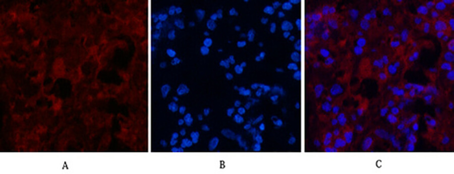
Fig.2. Immunofluorescence analysis of human appendix tissue. 1, MICU1 Monoclonal Antibody (red) was diluted at 1:200 (4°C, overnight). 2, Cy3 Labeled secondary antibody was diluted at 1:300 (room temperature, 50min). 3, Picture B: DAPI (blue) 10min. Picture A: Target. Picture B: DAPI. Picture C: merge of A+B.
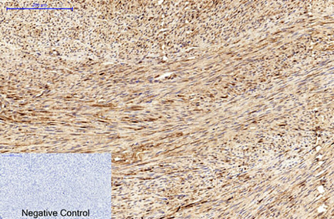
Fig.3. Immunohistochemical analysis of paraffin-embedded human uterus tissue. 1, MICU1 Monoclonal Antibody was diluted at 1:200 (4°C, overnight). 2, Sodium citrate pH 6.0 was used for antibody retrieval (>98°C, 20min). 3, secondary antibody was diluted at 1:200 (room temperature, 30min). Negative control was used by secondary antibody only.
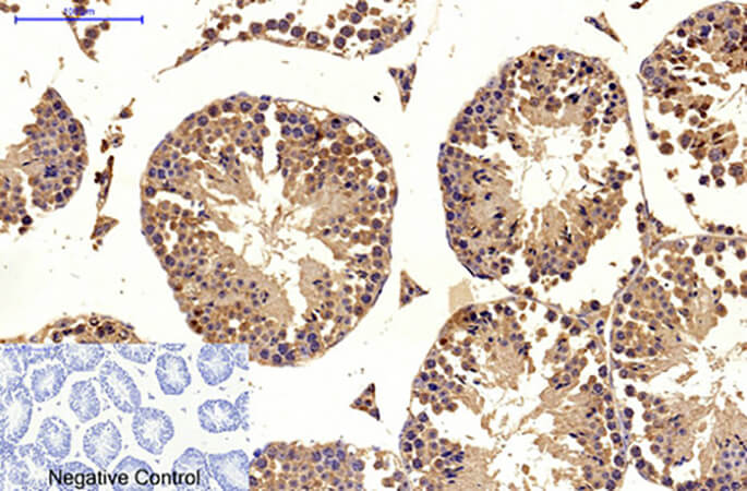
Fig.4. Immunohistochemical analysis of paraffin-embedded mouse testis tissue. 1, MICU1 Monoclonal Antibody was diluted at 1:200 (4°C, overnight). 2, Sodium citrate pH 6.0 was used for antibody retrieval (>98°C, 20min). 3, secondary antibody was diluted at 1:200 (room temperature, 30min). Negative control was used by secondary antibody only.
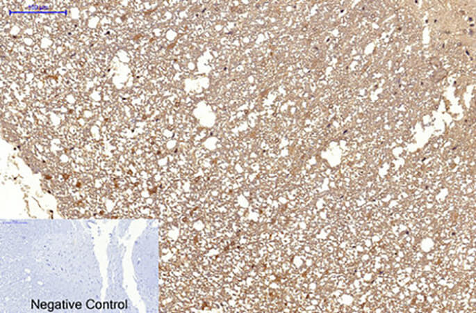
Fig.5. Immunohistochemical analysis of paraffin-embedded rat spinal cord tissue. 1, MICU1 Monoclonal Antibody was diluted at 1:200 (4°C, overnight). 2, Sodium citrate pH 6.0 was used for antibody retrieval (>98°C, 20min). 3, secondary antibody was diluted at 1:200 (room temperature, 30min). Negative control was used by secondary antibody only.
You must be logged in to post a review.


Reviews
There are no reviews yet.