| Product name | Luciferase Mouse Monoclonal Antibody (6B8) |
| Immunogen | Recombinant Protein of Luciferase of Luciferase |
| Host | Mouse |
| Reactivity | Firefly |
| Applications | IF,WB, |
| Applications notes | Optimal working dilutions should be determined experimentally by the investigator. Suggested starting dilutions are as follows: IF 1:50-200;WB 1:1000-2000 |
| Clonality | Monoclonal |
| Preparation method | The antibody was affinity-purified from mouse ascites by affinity-chromatography using specific immunogen. |
| Formulation | Liquid solution |
| Concentration | 1 mg/ml |
| Molecular weight | 60kD |
| Storage buffer | Liquid in PBS containing 50% glycerol, 0.5% BSA and 0.02% sodium azide. |
| Storage instructions | Stable for one year at -20°C from date of shipment. For maximum recovery of product, centrifuge the original vial after thawing and prior to removing the cap. Aliquot to avoid repeated freezing and thawing. |
| Shipping | Gel pack with blue ice. |
| Precautions | The product listed herein is for research use only and is not intended for use in human or clinical diagnosis. Suggested applications of our products are not recommendations to use our products in violation of any patent or as a license. We cannot be responsible for patent infringements or other violations that may occur with the use of this product. |
| Background | Luciferase from the firefly has become one of the more widely used reporter proteins for the study of gene expression. Luciferase catalyzes a bioluminescent reaction which requires the substrate luciferin as well as Mg2+ and ATP. Mixing these reagents with the cell extract containing luciferase, results in a flash of light that decays rapidly. This light can be detected by a luminometer. The total light emission is proportional to the luciferase activity of the sample. |
| Gene ID | N/A |
| Others | Luciferase protein detects endogenous levels of Luciferase. |
| Accession | P08659 |
| Observed Band(KD) | 60 |

Fig.1. Immunofluorescence analysis of human lung tissue. 1, Luciferase Mouse Monoclonal Antibody (6B8) (red) was diluted at 1:200 (4°C, overnight). 2, Cy3 Labeled secondary antibody was diluted at 1:300 (room temperature, 50min). 3, Picture B: DAPI (blue) 10min. Picture A: Target. Picture B: DAPI. Picture C: merge of A+B.
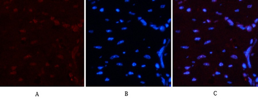
Fig.2. Immunofluorescence analysis of mouse heart tissue. 1, Luciferase Mouse Monoclonal Antibody (6B8) (red) was diluted at 1:200 (4°C, overnight). 2, Cy3 Labeled secondary antibody was diluted at 1:300 (room temperature, 50min). 3, Picture B: DAPI (blue) 10min. Picture A: Target. Picture B: DAPI. Picture C: merge of A+B.

Fig.3. Immunohistochemical analysis of paraffin-embedded rat lung tissue. 1, Luciferase Mouse Monoclonal Antibody (6B8) was diluted at 1:200 (4°C, overnight). 2, Sodium citrate pH 6.0 was used for antibody retrieval (>98°C, 20min). 3, secondary antibody was diluted at 1:200 (room temperature, 30min). Negative control was used by secondary antibody only.
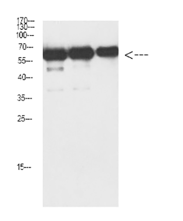
Fig.4. Western Blot analysis of Luciferase protein using antibody diluted at 1:1000.
You must be logged in to post a review.


Reviews
There are no reviews yet.