| Product name | FAK Polyclonal Antibody |
| Immunogen | Synthesized peptide derived from human FAK around the non-phosphorylation site of S843 |
| Host | Rabbit |
| Reactivity | Human,Mouse,Rat |
| Applications | IF,WB,IHC,ELISA |
| Applications notes | Optimal working dilutions should be determined experimentally by the investigator. Suggested starting dilutions are as follows: IF 1:50-200;WB 1:500-1:2000;IHC 1:100-1:300;ELISA 1:40000;Not yet tested in other applications; |
| Clonality | Polyclonal |
| Preparation method | The antibody was affinity-purified from rabbit antiserum by affinity-chromatography using epitope-specific immunogen. |
| Alternative | PTK2; FAK; FAK1; Focal adhesion kinase 1; FADK 1; Focal adhesion kinase-related nonkinase; FRNK; Protein phosphatase 1 regulatory subunit 71; PPP1R71; Protein-tyrosine kinase 2; p125FAK; pp125FAK |
| Formulation | Liquid solution |
| Concentration | 1 mg/ml |
| Molecular weight | 120kD |
| Storage buffer | Liquid in PBS containing 50% glycerol, 0.5% BSA and 0.02% sodium azide. |
| Storage instructions | Stable for one year at -20°C from date of shipment. For maximum recovery of product, centrifuge the original vial after thawing and prior to removing the cap. Aliquot to avoid repeated freezing and thawing. |
| Shipping | Gel pack with blue ice. |
| Precautions | The product listed herein is for research use only and is not intended for use in human or clinical diagnosis. Suggested applications of our products are not recommendations to use our products in violation of any patent or as a license. We cannot be responsible for patent infringements or other violations that may occur with the use of this product. |
| Background | PTK2 (protein tyrosine kinase 2) encodes a cytoplasmic protein tyrosine kinase which is found concentrated in the focal adhesions that form between cells growing in the presence of extracellular matrix constituents. The encoded protein is a member of the FAK subfamily of protein tyrosine kinases but lacks significant sequence similarity to kinases from other subfamilies. Activation of PTK2 may be an important early step in cell growth and intracellular signal transduction pathways triggered in response to certain neural peptides or to cell interactions with the extracellular matrix. Several transcript variants encoding different isoforms have been found for this gene, but the full-length natures of only four of them have been determined. |
| Gene ID | 5747 |
| Alternative | PTK2; FAK; FAK1; Focal adhesion kinase 1; FADK 1; Focal adhesion kinase-related nonkinase; FRNK; Protein phosphatase 1 regulatory subunit 71; PPP1R71; Protein-tyrosine kinase 2; p125FAK; pp125FAK |
| Others | FAK Polyclonal Antibody detects endogenous levels of FAK protein. |
| Accession | Q05397 |
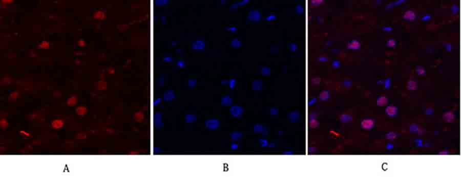
Fig.1. Immunofluorescence analysis of human liver tissue. 1, FAK Polyclonal Antibody (red) was diluted at 1:200 (4°C, overnight). 2, Cy3 Labeled secondary antibody was diluted at 1:300 (room temperature, 50min). 3, Picture B: DAPI (blue) 10min. Picture A: Target. Picture B: DAPI. Picture C: merge of A+B.
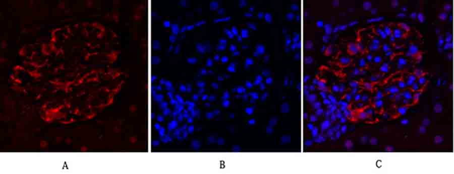
Fig.2. Immunofluorescence analysis of rat kidney tissue. 1, FAK Polyclonal Antibody (red) was diluted at 1:200 (4°C, overnight). 2, Cy3 Labeled secondary antibody was diluted at 1:300 (room temperature, 50min). 3, Picture B: DAPI (blue) 10min. Picture A: Target. Picture B: DAPI. Picture C: merge of A+B.
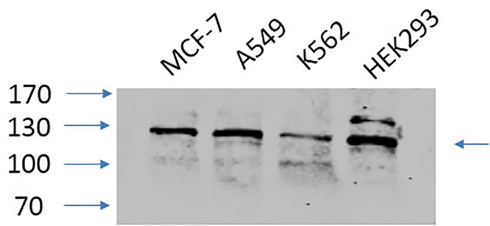
Fig.3. Western Blot analysis of MCF-7 (1), A549 (2), K562 (3), HEK293 (4), diluted at 1:1000.
You must be logged in to post a review.


Reviews
There are no reviews yet.