| Product name | ERK 1/2 Polyclonal Antibody |
| Immunogen | Synthesized peptide derived from the C-terminal region of human ERK 1/2 at AA range: 300-380 |
| Host | Rabbit |
| Reactivity | Human,Mouse,Rat |
| Applications | IF,WB,IHC,ELISA,ChIP |
| Applications notes | Optimal working dilutions should be determined experimentally by the investigator. Suggested starting dilutions are as follows: IF 1:50-200;WB 1:500-1:2000;IHC 1:100-1:300;ELISA 1:10000;Not yet tested in other applications; |
| Clonality | Polyclonal |
| Preparation method | The antibody was affinity-purified from rabbit antiserum by affinity-chromatography using epitope-specific immunogen. |
| Alternative | MAPK3; ERK1; PRKM3; Mitogen-activated protein kinase 3; MAP kinase 3; MAPK 3; ERT2; Extracellular signal-regulated kinase 1; ERK-1; Insulin-stimulated MAP2 kinase; MAP kinase isoform p44; p44-MAPK; Microtubule-associated protein 2 kinase; p |
| Formulation | Liquid solution |
| Concentration | 1 mg/ml |
| Molecular weight | 42,44kD |
| Storage buffer | Liquid in PBS containing 50% glycerol, 0.5% BSA and 0.02% sodium azide. |
| Storage instructions | Stable for one year at -20°C from date of shipment. For maximum recovery of product, centrifuge the original vial after thawing and prior to removing the cap. Aliquot to avoid repeated freezing and thawing. |
| Shipping | Gel pack with blue ice. |
| Precautions | The product listed herein is for research use only and is not intended for use in human or clinical diagnosis. Suggested applications of our products are not recommendations to use our products in violation of any patent or as a license. We cannot be responsible for patent infringements or other violations that may occur with the use of this product. |
| Background | Mitogen-activated protein kinase 3 encoded by MAPK3 is a member of the MAP kinase family. MAP kinases, also known as extracellular signal-regulated kinases (ERKs), act in a signaling cascade that regulates various cellular processes such as proliferation, differentiation, and cell cycle progression in response to a variety of extracellular signals. This kinase is activated by upstream kinases, resulting in its translocation to the nucleus where it phosphorylates nuclear targets. Alternatively spliced transcript variants encoding different protein isoforms have been described. |
| Gene ID | 5595 |
| Alternative | MAPK3; ERK1; PRKM3; Mitogen-activated protein kinase 3; MAP kinase 3; MAPK 3; ERT2; Extracellular signal-regulated kinase 1; ERK-1; Insulin-stimulated MAP2 kinase; MAP kinase isoform p44; p44-MAPK; Microtubule-associated protein 2 kinase; p |
| Others | ERK 1/2 Polyclonal Antibody detects endogenous levels of ERK 1/2 protein. |
| Accession | P27361/P28482 |
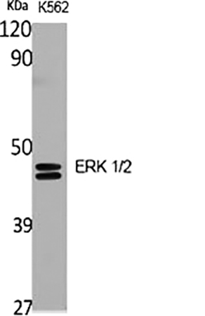
Fig.1. Western Blot analysis of various cells using ERK 1/2 Polyclonal Antibody diluted at 1:2000.
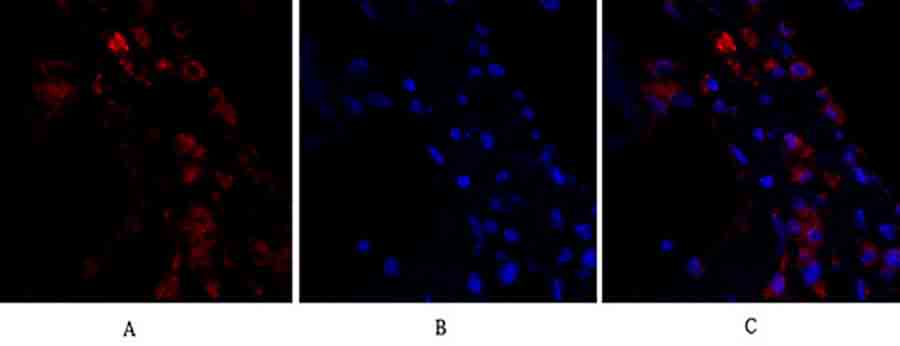
Fig.2. Immunofluorescence analysis of human lung tissue. 1, ERK 1/2 Polyclonal Antibody (red) was diluted at 1:200 (4°C, overnight). 2, Cy3 Labeled secondary antibody was diluted at 1:300 (room temperature, 50min). 3, Picture B: DAPI (blue) 10min. Picture A: Target. Picture B: DAPI. Picture C: merge of A+B.
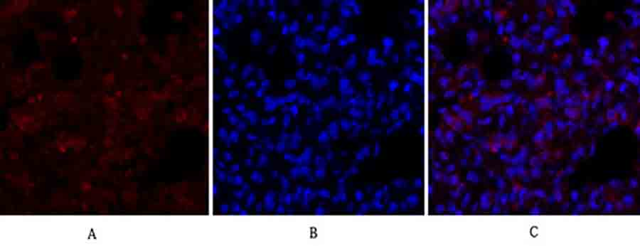
Fig.3. Immunofluorescence analysis of rat lung tissue. 1, ERK 1/2 Polyclonal Antibody (red) was diluted at 1:200 (4°C, overnight). 2, Cy3 Labeled secondary antibody was diluted at 1:300 (room temperature, 50min). 3, Picture B: DAPI (blue) 10min. Picture A: Target. Picture B: DAPI. Picture C: merge of A+B.
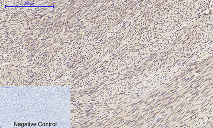
Fig.4. Immunohistochemical analysis of paraffin-embedded human uterus tissue. 1, ERK 1/2 Polyclonal Antibody was diluted at 1:200 (4°C, overnight). 2, Sodium citrate pH 6.0 was used for antibody retrieval (>98°C, 20min). 3, secondary antibody was diluted at 1:200 (room temperature, 30min). Negative control was used by secondary antibody only.
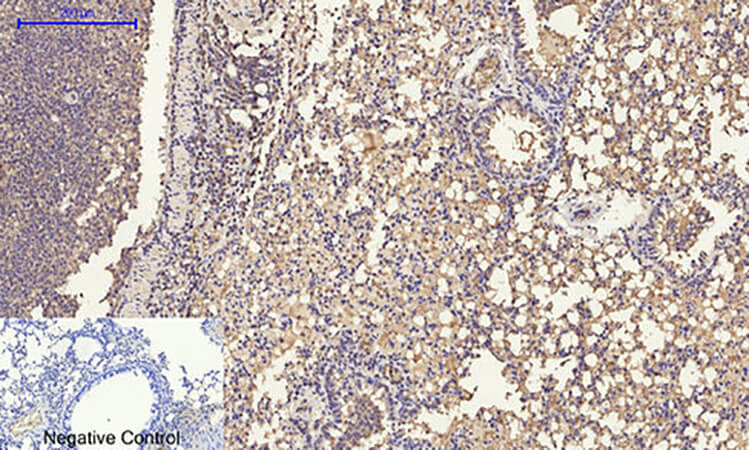
Fig.5. Immunohistochemical analysis of paraffin-embedded mouse lung tissue. 1, ERK 1/2 Polyclonal Antibody was diluted at 1:200 (4°C, overnight). 2, Sodium citrate pH 6.0 was used for antibody retrieval (>98°C, 20min). 3, secondary antibody was diluted at 1:200 (room temperature, 30min). Negative control was used by secondary antibody only.
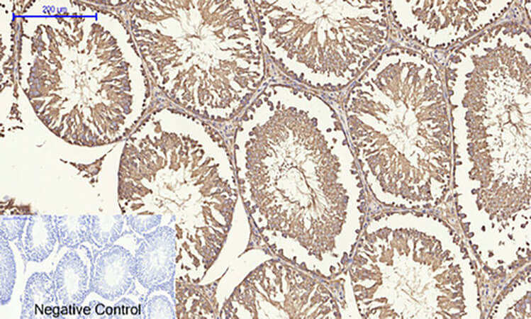
Fig.6. Immunohistochemical analysis of paraffin-embedded rat testis tissue. 1, ERK 1/2 Polyclonal Antibody was diluted at 1:200 (4°C, overnight). 2, Sodium citrate pH 6.0 was used for antibody retrieval (>98°C, 20min). 3, secondary antibody was diluted at 1:200 (room temperature, 30min). Negative control was used by secondary antibody only.
Author:Chang, Chenli, et al Publication name:Respiratory Research IF:6
Author:Liu W, Wang L, Zhang J, Qiao L, Liu Y, Yang X, Zhang J, Zheng W, Ma Z Publication name:3 Biotech IF:2
You must be logged in to post a review.


Reviews
There are no reviews yet.