| Product name | E-cadherin Polyclonal Antibody |
| Immunogen | Synthesized peptide derived from the C-terminal region of human E-cadherin at AA range: 810-890 |
| Host | Rabbit |
| Reactivity | Human,Mouse,Rat |
| Applications | WB,IHC,IF,ELISA |
| Applications notes | Optimal working dilutions should be determined experimentally by the investigator. Suggested starting dilutions are as follows: WB 1:500-1:2000;IHC: 1:100-300;ELISA 1:20000;IF 1:100-300;Not yet tested in other applications. |
| Clonality | Polyclonal |
| Preparation method | The antibody was affinity-purified from rabbit antiserum by affinity-chromatography using epitope-specific immunogen. |
| Alternative | CDH1; CDHE; UVO; Cadherin-1; CAM 120/80; Epithelial cadherin; E-cadherin; Uvomorulin; CD antigen CD324; CDH2; CDHN; NCAD; Cadherin-2; CDw325; Neural cadherin; N-cadherin; CD antigen CD325; CDH3; CDHP; Cadherin-3; Placental cadherin; P-cadhe |
| Formulation | Liquid solution |
| Concentration | 1 mg/ml |
| Molecular weight | 125-130kD |
| Storage buffer | Liquid in PBS containing 50% glycerol, 0.5% BSA and 0.02% sodium azide. |
| Storage instructions | Stable for one year at -20°C from date of shipment. For maximum recovery of product, centrifuge the original vial after thawing and prior to removing the cap. Aliquot to avoid repeated freezing and thawing. |
| Shipping | Gel pack with blue ice. |
| Precautions | The product listed herein is for research use only and is not intended for use in human or clinical diagnosis. Suggested applications of our products are not recommendations to use our products in violation of any patent or as a license. We cannot be responsible for patent infringements or other violations that may occur with the use of this product. |
| Background | CDH1 encodes a classical cadherin of the cadherin superfamily. Alternative splicing results in multiple transcript variants, at least one of which encodes a preproprotein that is proteolytically processed to generate the mature glycoprotein. This calcium-dependent cell-cell adhesion protein is comprised of five extracellular cadherin repeats, a transmembrane region and a highly conserved cytoplasmic tail. Mutations in CDH1 are correlated with gastric, breast, colorectal, thyroid and ovarian cancer. Loss of function of this gene is thought to contribute to cancer progression by increasing proliferation, invasion, and/or metastasis. The ectodomain of this protein mediates bacterial adhesion to mammalian cells and the cytoplasmic domain is required for internalization. CDH1 is present in a gene cluster with other members of the cadherin family on chromosome 16. |
| Gene ID | 999/1000/1001/1002 |
| Alternative | CDH1; CDHE; UVO; Cadherin-1; CAM 120/80; Epithelial cadherin; E-cadherin; Uvomorulin; CD antigen CD324; CDH2; CDHN; NCAD; Cadherin-2; CDw325; Neural cadherin; N-cadherin; CD antigen CD325; CDH3; CDHP; Cadherin-3; Placental cadherin; P-cadhe |
| Others | E-cadherin Polyclonal Antibody detects endogenous levels of E-cadherin protein. |
| Accession | P12830/P19022/P22223/P55283 |

Fig.1. Western Blot analysis of various cells using E-cadherin Polyclonal Antibody diluted at 1:2000.
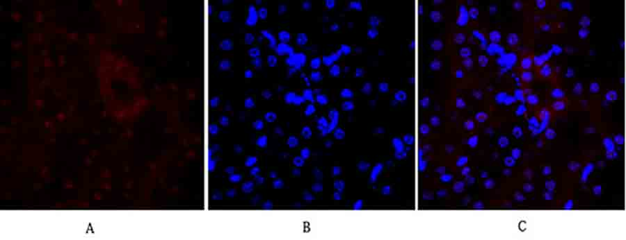
Fig.2. Immunofluorescence analysis of mouse kidney tissue. 1, E-cadherin Polyclonal Antibody (red) was diluted at 1:200 (4°C, overnight). 2, Cy3 labeled secondary antibody was diluted at 1:300 (room temperature, 50min). 3, Picture B: DAPI (blue) 10min. Picture A: Target. Picture B: DAPI. Picture C: merge of A+B.
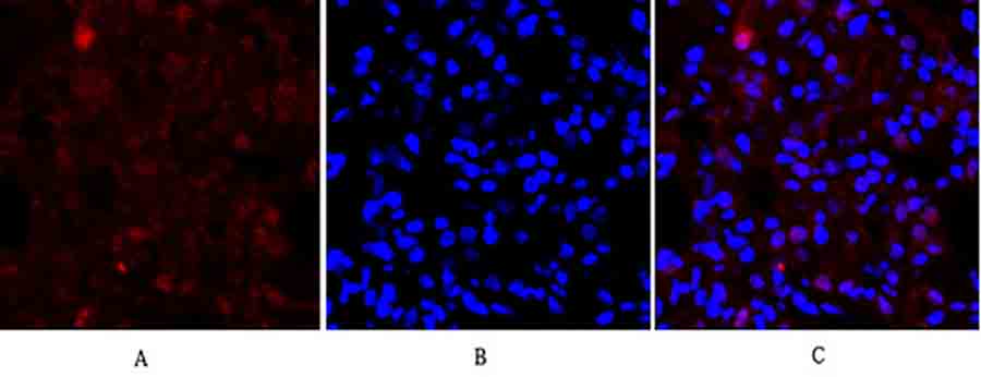
Fig.3. Immunofluorescence analysis of rat lung tissue. 1, E-cadherin Polyclonal Antibody (red) was diluted at 1:200 (4°C, overnight). 2, Cy3 labeled secondary antibody was diluted at 1:300 (room temperature, 50min). 3, Picture B: DAPI (blue) 10min. Picture A: Target. Picture B: DAPI. Picture C: merge of A+B.
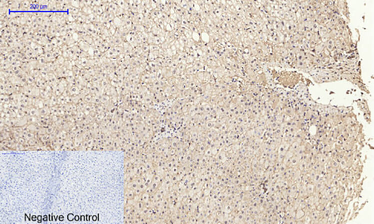
Fig.4. Immunohistochemical analysis of paraffin-embedded human liver tissue. 1, E-cadherin Polyclonal Antibody was diluted at 1:200 (4°C, overnight). 2, Sodium citrate pH 6.0 was used for antibody retrieval (>98°C, 20min). 3, secondary antibody was diluted at 1:200 (room temperature, 30min). Negative control was used by secondary antibody only.
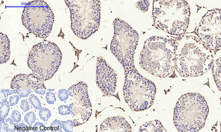
Fig.5. Immunohistochemical analysis of paraffin-embedded mouse testis tissue. 1, E-cadherin Polyclonal Antibody was diluted at 1:200 (4°C, overnight). 2, Sodium citrate pH 6.0 was used for antibody retrieval (>98°C, 20min). 3, secondary antibody was diluted at 1:200 (room temperature, 30min). Negative control was used by secondary antibody only.
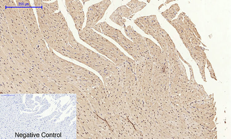
Fig.6. Immunohistochemical analysis of paraffin-embedded rat heart tissue. 1, E-cadherin Polyclonal Antibody was diluted at 1:200 (4°C, overnight). 2, Sodium citrate pH 6.0 was used for antibody retrieval (>98°C, 20min). 3, secondary antibody was diluted at 1:200 (room temperature, 30min). Negative control was used by secondary antibody only.
Author:Hossain AJ, Islam R, Kim JG Publication name:Biomedicines . IF:5
Author:W Liang, J Liu, H Wu, X Qiao, X Lu Publication name:Oncology letters IF:2
You must be logged in to post a review.


Reviews
There are no reviews yet.