| Product name | Cyclin D1 Polyclonal Antibody |
| Immunogen | Synthesized peptide derived from human Cyclin D1 around the non-phosphorylation site of T286 |
| Host | Rabbit |
| Reactivity | Human,Mouse,Rat,Pig |
| Applications | IF,WB,IHC,ELISA |
| Applications notes | Optimal working dilutions should be determined experimentally by the investigator. Suggested starting dilutions are as follows: IF 1:50-200;WB 1:500-1:2000;IHC 1:100-1:300;ELISA 1:40000;Not yet tested in other applications; |
| Clonality | Polyclonal |
| Preparation method | The antibody was affinity-purified from rabbit antiserum by affinity-chromatography using epitope-specific immunogen. |
| Alternative | CCND1; BCL1; PRAD1; G1/S-specific cyclin-D1; B-cell lymphoma 1 protein; BCL-1; BCL-1 oncogene; PRAD1 oncogene |
| Formulation | Liquid solution |
| Concentration | 1 mg/ml |
| Molecular weight | 33kD |
| Storage buffer | Liquid in PBS containing 50% glycerol, 0.5% BSA and 0.02% sodium azide. |
| Storage instructions | Stable for one year at -20°C from date of shipment. For maximum recovery of product, centrifuge the original vial after thawing and prior to removing the cap. Aliquot to avoid repeated freezing and thawing. |
| Shipping | Gel pack with blue ice. |
| Precautions | The product listed herein is for research use only and is not intended for use in human or clinical diagnosis. Suggested applications of our products are not recommendations to use our products in violation of any patent or as a license. We cannot be responsible for patent infringements or other violations that may occur with the use of this product. |
| Background | Cyclin D1 encoded by CCND1 belongs to the highly conserved cyclin family, whose members are characterized by a dramatic periodicity in protein abundance throughout the cell cycle. Cyclins function as regulators of CDK kinases. Different cyclins exhibit distinct expression and degradation patterns which contribute to the temporal coordination of each mitotic event. This cyclin forms a complex with and functions as a regulatory subunit of CDK4 or CDK6, whose activity is required for cell cycle G1/S transition. Cyclin D1 has been shown to interact with tumor suppressor protein Rb and the expression of this gene is regulated positively by Rb. Mutations, amplification and overexpression of CCND1, which alters cell cycle progression, are observed frequently in a variety of tumors and may contribute to tumorigenesis. |
| Gene ID | 595 |
| Alternative | CCND1; BCL1; PRAD1; G1/S-specific cyclin-D1; B-cell lymphoma 1 protein; BCL-1; BCL-1 oncogene; PRAD1 oncogene |
| Others | Cyclin D1 Polyclonal Antibody detects endogenous levels of Cyclin D1 protein. |
| Accession | P24385 |
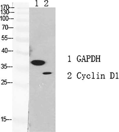
Fig.1. Western Blot analysis of GAPDH (1), Cyclin D1 (2), diluted at 1:1000.
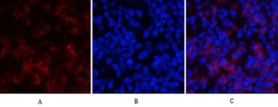
Fig.2. Immunofluorescence analysis of mouse spleen tissue. 1, Cyclin D1 Polyclonal Antibody (red) was diluted at 1:200 (4°C, overnight). 2, Cy3 labeled secondary antibody was diluted at 1:300 (room temperature, 50min). 3, Picture B: DAPI (blue) 10min. Picture A: Target. Picture B: DAPI. Picture C: merge of A+B.
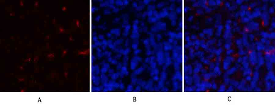
Fig.3. Immunofluorescence analysis of rat spleen tissue. 1, Cyclin D1 Polyclonal Antibody (red) was diluted at 1:200 (4°C, overnight). 2, Cy3 labeled secondary antibody was diluted at 1:300 (room temperature, 50min). 3, Picture B: DAPI (blue) 10min. Picture A: Target. Picture B: DAPI. Picture C: merge of A+B.
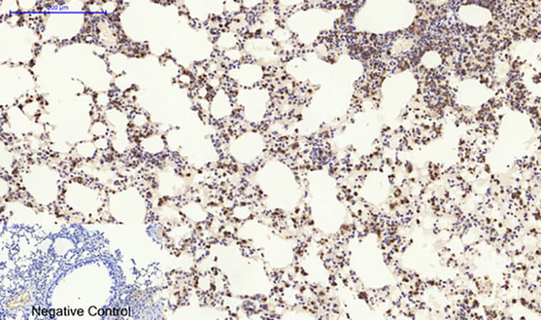
Fig.4. Immunohistochemical analysis of paraffin-embedded mouse lung tissue. 1, Cyclin D1 Polyclonal Antibody was diluted at 1:200 (4°C, overnight). 2, Sodium citrate pH 6.0 was used for antibody retrieval (>98°C, 20min). 3, secondary antibody was diluted at 1:200 (room temperature, 30min). Negative control was used by secondary antibody only.
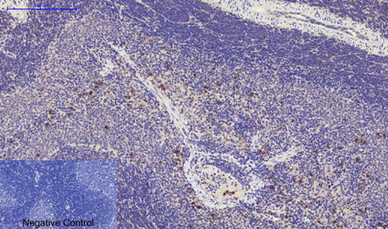
Fig.5. Immunohistochemical analysis of paraffin-embedded rat spleen tissue. 1, Cyclin D1 Polyclonal Antibody was diluted at 1:200 (4°C, overnight). 2, Sodium citrate pH 6.0 was used for antibody retrieval (>98°C, 20min). 3, secondary antibody was diluted at 1:200 (room temperature, 30min). Negative control was used by secondary antibody only.
You must be logged in to post a review.


Reviews
There are no reviews yet.