| Product name | Cyclin A Polyclonal Antibody |
| Immunogen | Synthesized peptide derived from the Internal region of human Cyclin A at AA range: 190-270 |
| Host | Rabbit |
| Reactivity | Human,Mouse,Rat,Monkey |
| Applications | WB,IHC,IF,ELISA |
| Applications notes | Optimal working dilutions should be determined experimentally by the investigator. Suggested starting dilutions are as follows: WB 1:500-1:2000;IHC 1:100-1:300;IF 1:200-1:1000;ELISA 1:10000;Not yet tested in other applications. |
| Clonality | Polyclonal |
| Preparation method | The antibody was affinity-purified from rabbit antiserum by affinity-chromatography using epitope-specific immunogen. |
| Alternative | CCNA1; Cyclin-A1; CCNA2; CCN1; CCNA; Cyclin-A2; Cyclin-A |
| Formulation | Liquid solution |
| Concentration | 1 mg/ml |
| Molecular weight | 53kD |
| Storage buffer | Liquid in PBS containing 50% glycerol, 0.5% BSA and 0.02% sodium azide. |
| Storage instructions | Stable for one year at -20°C from date of shipment. For maximum recovery of product, centrifuge the original vial after thawing and prior to removing the cap. Aliquot to avoid repeated freezing and thawing. |
| Shipping | Gel pack with blue ice. |
| Precautions | The product listed herein is for research use only and is not intended for use in human or clinical diagnosis. Suggested applications of our products are not recommendations to use our products in violation of any patent or as a license. We cannot be responsible for patent infringements or other violations that may occur with the use of this product. |
| Background | Cyclin A1 encoded by CCNA1 belongs to the highly conserved cyclin family, whose members are characterized by a dramatic periodicity in protein abundance through the cell cycle. Cyclins function as regulators of CDK kinases. Different cyclins exhibit distinct expression and degradation patterns which contribute to the temporal coordination of each mitotic event. The cyclin encoded by this gene was shown to be expressed in testis and brain, as well as in several leukemic cell lines, and is thought to primarily function in the control of the germline meiotic cell cycle. This cyclin binds both CDK2 and CDC2 kinases, which give two distinct kinase activities, one appearing in S phase, the other in G2, and thus regulate separate functions in cell cycle. This cyclin was found to bind to important cell cycle regulators, such as Rb family proteins, transcription factor E2F-1, and the p21 family proteins. Multiple transcript variants encoding different isoforms have been found for CCNA1. |
| Gene ID | 8900/890 |
| Alternative | CCNA1; Cyclin-A1; CCNA2; CCN1; CCNA; Cyclin-A2; Cyclin-A |
| Others | Cyclin A Polyclonal Antibody detects endogenous levels of Cyclin A protein. |
| Accession | P78396/P20248 |
| Observed Band(KD) | 53 |
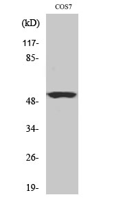
Fig.1. Western Blot analysis of COS7 cells using Cyclin A Polyclonal Antibody, diluted at 1: 500.
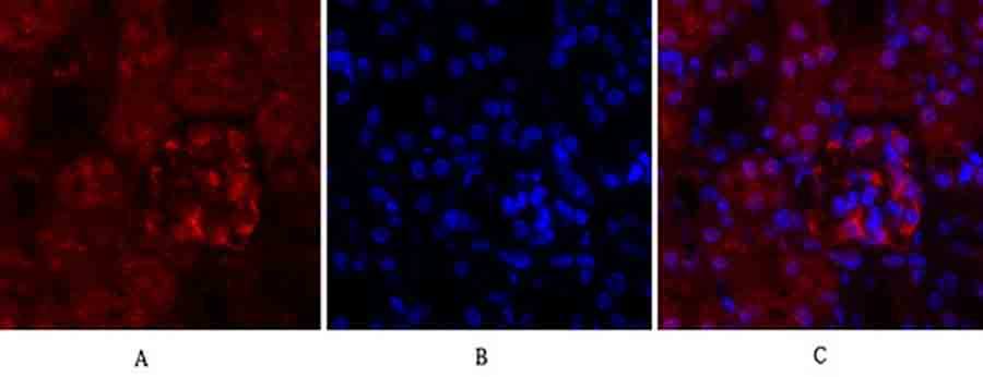
Fig.2. Immunofluorescence analysis of mouse kidney tissue. 1, Cyclin A Polyclonal Antibody (red) was diluted at 1:200 (4°C, overnight). 2, Cy3 labeled secondary antibody was diluted at 1:300 (room temperature, 50min). 3, Picture B: DAPI (blue) 10min. Picture A: Target. Picture B: DAPI. Picture C: merge of A+B.
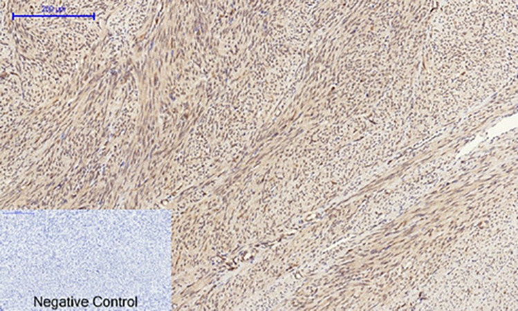
Fig.3. Immunohistochemical analysis of paraffin-embedded human uterus tissue. 1, Cyclin A Polyclonal Antibody was diluted at 1:200 (4°C, overnight). 2, Sodium citrate pH 6.0 was used for antibody retrieval (>98°C, 20min). 3, secondary antibody was diluted at 1:200 (room temperature, 30min). Negative control was used by secondary antibody only.
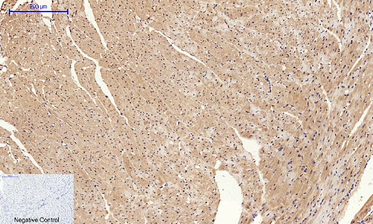
Fig.4. Immunohistochemical analysis of paraffin-embedded mouse heart tissue. 1, Cyclin A Polyclonal Antibody was diluted at 1:200 (4°C, overnight). 2, Sodium citrate pH 6.0 was used for antibody retrieval (>98°C, 20min). 3, secondary antibody was diluted at 1:200 (room temperature, 30min). Negative control was used by secondary antibody only.
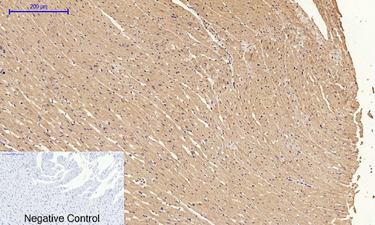
Fig.5. Immunohistochemical analysis of paraffin-embedded rat heart tissue. 1, Cyclin A Polyclonal Antibody was diluted at 1:200 (4°C, overnight). 2, Sodium citrate pH 6.0 was used for antibody retrieval (>98°C, 20min). 3, secondary antibody was diluted at 1:200 (room temperature, 30min). Negative control was used by secondary antibody only.
You must be logged in to post a review.


Reviews
There are no reviews yet.