| Product name | Cleaved-PARP-1 (D214) Polyclonal Antibody |
| Immunogen | Synthesized peptide derived from the Internal region of human PARP-1 at AA range: 140-220 |
| Host | Rabbit |
| Reactivity | Human,Mouse |
| Applications | WB,IHC,IF,ELISA |
| Applications notes | Optimal working dilutions should be determined experimentally by the investigator. Suggested starting dilutions are as follows: WB 1:500-2000;IF 1:50-300;IHC 1:50-300 |
| Clonality | Polyclonal |
| Preparation method | The antibody was affinity-purified from rabbit antiserum by affinity-chromatography using epitope-specific immunogen. |
| Alternative | PARP1; ADPRT; PPOL; Poly [ADP-ribose] polymerase 1; PARP-1; ADP-ribosyltransferase diphtheria toxin-like 1; ARTD1; NAD(+) ADP-ribosyltransferase 1; ADPRT 1; Poly[ADP-ribose] synthase 1 |
| Formulation | Liquid solution |
| Concentration | 1 mg/ml |
| Molecular weight | 24kD |
| Storage buffer | Liquid in PBS containing 50% glycerol, 0.5% BSA and 0.02% sodium azide. |
| Storage instructions | Stable for one year at -20°C from date of shipment. For maximum recovery of product, centrifuge the original vial after thawing and prior to removing the cap. Aliquot to avoid repeated freezing and thawing. |
| Shipping | Gel pack with blue ice. |
| Precautions | The product listed herein is for research use only and is not intended for use in human or clinical diagnosis. Suggested applications of our products are not recommendations to use our products in violation of any patent or as a license. We cannot be responsible for patent infringements or other violations that may occur with the use of this product. |
| Background | PARP1 encodes a chromatin-associated enzyme, poly(ADP-ribosyl)transferase, which modifies various nuclear proteins by poly(ADP-ribosyl)ation. The modification is dependent on DNA and is involved in the regulation of various important cellular processes such as differentiation, proliferation, and tumor transformation and also in the regulation of the molecular events involved in the recovery of cell from DNA damage. In addition, poly(ADP-ribose) polymerase 1 may be the site of mutation in Fanconi anemia, and may participate in the pathophysiology of type I diabetes. |
| Gene ID | 142 |
| Alternative | PARP1; ADPRT; PPOL; Poly [ADP-ribose] polymerase 1; PARP-1; ADP-ribosyltransferase diphtheria toxin-like 1; ARTD1; NAD(+) ADP-ribosyltransferase 1; ADPRT 1; Poly[ADP-ribose] synthase 1 |
| Others | Cleaved-PARP-1 (D214) Polyclonal Antibody detects endogenous levels of fragment of activated PARP-1 protein resulting from cleavage adjacent to D214. |
| Accession | P09874 |
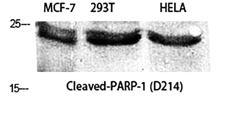
Fig.1. Western Blot analysis of MCF-7 (1), 293T (2), Hela (3), diluted at 1:2000.
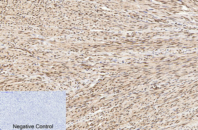
Fig.2. Immunohistochemical analysis of paraffin-embedded human uterus tissue. 1, Cleaved-PARP-1 (D214) Polyclonal Antibody was diluted at 1:200 (4°C, overnight). 2, Sodium citrate pH 6.0 was used for antibody retrieval (>98°C, 20min). 3, secondary antibody was diluted at 1:200 (room temperature, 30min). Negative control was used by secondary antibody only.
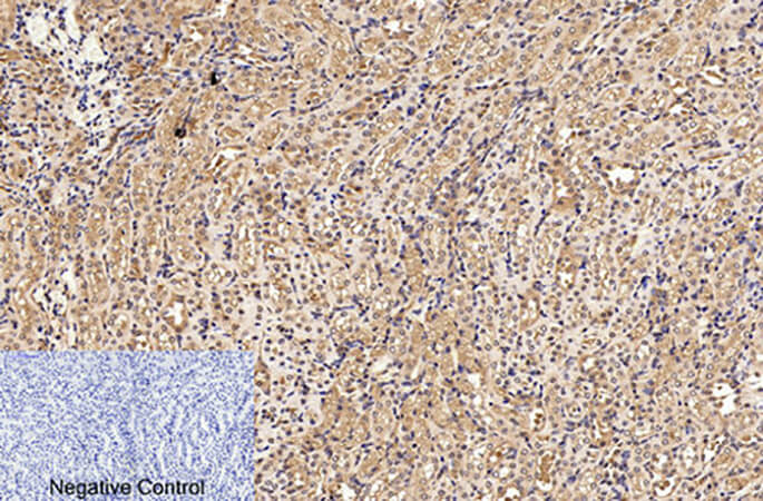
Fig.3. Immunohistochemical analysis of paraffin-embedded mouse kidney tissue. 1, Cleaved-PARP-1 (D214) Polyclonal Antibody was diluted at 1:200 (4°C, overnight). 2, Sodium citrate pH 6.0 was used for antibody retrieval (>98°C, 20min). 3, secondary antibody was diluted at 1:200 (room temperature, 30min). Negative control was used by secondary antibody only.
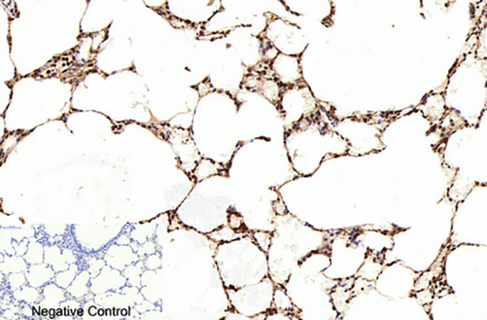
Fig.4. Immunohistochemical analysis of paraffin-embedded rat lung tissue. 1, Cleaved-PARP-1 (D214) Polyclonal Antibody was diluted at 1:200 (4°C, overnight). 2, Sodium citrate pH 6.0 was used for antibody retrieval (>98°C, 20min). 3, secondary antibody was diluted at 1:200 (room temperature, 30min). Negative control was used by secondary antibody only.
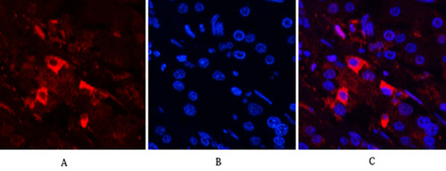
Fig.5. Immunofluorescence analysis of human stomach cancer tissue. 1, Cleaved-PARP-1 (D214) Polyclonal Antibody (red) was diluted at 1:200 (4°C, overnight). 2, Cy3 Labeled secondary antibody was diluted at 1:300 (room temperature, 50min). 3, Picture B: DAPI (blue) 10min. Picture A: Target. Picture B: DAPI. Picture C: merge of A+B.
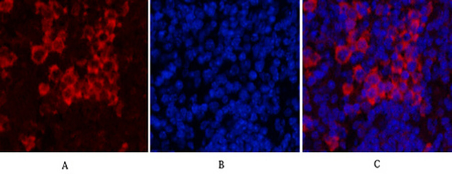
Fig.6. Immunofluorescence analysis of mouse spleen tissue. 1, Cleaved-PARP-1 (D214) Polyclonal Antibody (red) was diluted at 1:200 (4°C, overnight). 2, Cy3 Labeled secondary antibody was diluted at 1:300 (room temperature, 50min). 3, Picture B: DAPI (blue) 10min. Picture A: Target. Picture B: DAPI. Picture C: merge of A+B.
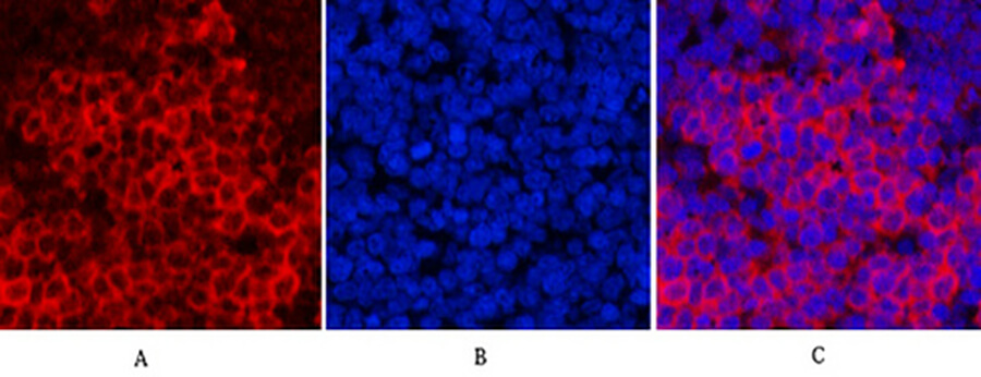
Fig.7. Immunofluorescence analysis of rat spleen tissue. 1, Cleaved-PARP-1 (D214) Polyclonal Antibody (red) was diluted at 1:200 (4°C, overnight). 2, Cy3 Labeled secondary antibody was diluted at 1:300 (room temperature, 50min). 3, Picture B: DAPI (blue) 10min. Picture A: Target. Picture B: DAPI. Picture C: merge of A+B.
You must be logged in to post a review.


Reviews
There are no reviews yet.