| Product name | CDX2 Monoclonal Antibody |
| Immunogen | Synthetic Peptide |
| Host | Mouse |
| Reactivity | Human,Mouse,Rat |
| Applications | WB,IF,IHC |
| Applications notes | Optimal working dilutions should be determined experimentally by the investigator. Suggested starting dilutions are as follows: WB 1:1000;IHC 1:200;IF 1:200 |
| Clonality | Monoclonal |
| Preparation method | The antibody was affinity-purified from mouse ascites by affinity-chromatography using specific immunogen. |
| Alternative | CDX2; CDX3; Homeobox protein CDX-2; CDX-3; Caudal-type homeobox protein 2 |
| Formulation | Liquid solution |
| Concentration | 1 mg/ml |
| Molecular weight | 42kD |
| Storage buffer | PBS, pH 7.4, containing 0.5%BSA, 0.02% sodium azide as Preservative and 50% Glycerol. |
| Storage instructions | Stable for one year at -20°C from date of shipment. For maximum recovery of product, centrifuge the original vial after thawing and prior to removing the cap. Aliquot to avoid repeated freezing and thawing. |
| Shipping | Gel pack with blue ice. |
| Precautions | The product listed herein is for research use only and is not intended for use in human or clinical diagnosis. Suggested applications of our products are not recommendations to use our products in violation of any patent or as a license. We cannot be responsible for patent infringements or other violations that may occur with the use of this product. |
| Background | CDX2 is a member of the caudal-related homeobox transcription factor gene family. caudal type homeobox 2 is a major regulator of intestine-specific genes involved in cell growth an differentiation. Caudal type homeobox 2 also plays a role in early embryonic development of the intestinal tract. Aberrant expression of CDX2 is associated with intestinal inflammation and tumorigenesis. |
| Gene ID | 1045 |
| Alternative | CDX2; CDX3; Homeobox protein CDX-2; CDX-3; Caudal-type homeobox protein 2 |
| Others | The antibody detects endogenous CDX2 proteins. |
| Accession | Q99626 |
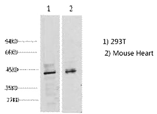
Fig.1. Western blot analysis of 1) 293T, 2) Mouse Heart tissue, diluted at 1:2000.
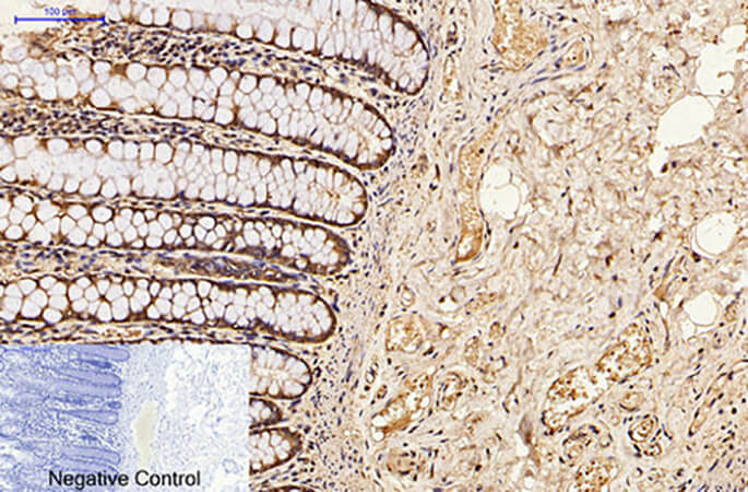
Fig.2. Immunohistochemical analysis of paraffin-embedded human colon tissue. 1, CDX2 Monoclonal Antibody was diluted at 1:200 (4°C, overnight). 2, Sodium citrate pH 6.0 was used for antibody retrieval (>98°C, 20min). 3, secondary antibody was diluted at 1:200 (room temperature, 30min). Negative control was used by secondary antibody only.
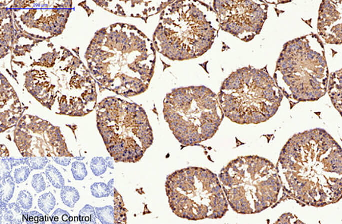
Fig.3. Immunohistochemical analysis of paraffin-embedded mouse testis tissue. 1, CDX2 Monoclonal Antibody was diluted at 1:200 (4°C, overnight). 2, Sodium citrate pH 6.0 was used for antibody retrieval (>98°C, 20min). 3, secondary antibody was diluted at 1:200 (room temperature, 30min). Negative control was used by secondary antibody only.
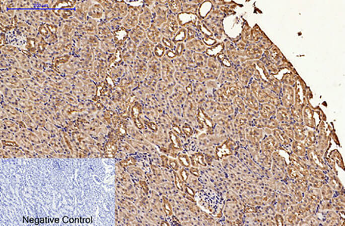
Fig.4. Immunohistochemical analysis of paraffin-embedded rat kidney tissue. 1, CDX2 Monoclonal Antibody was diluted at 1:200 (4°C, overnight). 2, Sodium citrate pH 6.0 was used for antibody retrieval (>98°C, 20min). 3, secondary antibody was diluted at 1:200 (room temperature, 30min). Negative control was used by secondary antibody only.
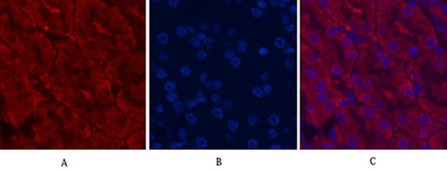
Fig.5. Immunofluorescence analysis of mouse kidney tissue. 1, CDX2 Monoclonal Antibody (red) was diluted at 1:200 (4°C, overnight). 2, Cy3 Labeled secondary antibody was diluted at 1:300 (room temperature, 50min). 3, Picture B: DAPI (blue) 10min. Picture A: Target. Picture B: DAPI. Picture C: merge of A+B.
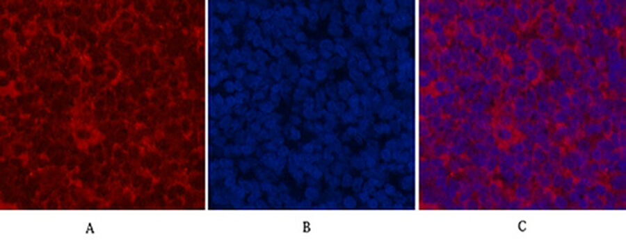
Fig.6. Immunofluorescence analysis of rat spleen tissue. 1, CDX2 Monoclonal Antibody (red) was diluted at 1:200 (4°C, overnight). 2, Cy3 Labeled secondary antibody was diluted at 1:300 (room temperature, 50min). 3, Picture B: DAPI (blue) 10min. Picture A: Target. Picture B: DAPI. Picture C: merge of A+B.
You must be logged in to post a review.


Reviews
There are no reviews yet.