| Product name | Cdk4 Polyclonal Antibody |
| Immunogen | Synthesized peptide derived from the N-terminal region of human Cdk4 |
| Host | Rabbit |
| Reactivity | Human,Mouse,Rat |
| Applications | IF,WB,IHC,ELISA |
| Applications notes | Optimal working dilutions should be determined experimentally by the investigator. Suggested starting dilutions are as follows: IF 1:50-200;WB 1:500-1:2000;IHC: 1:100-300;ELISA 1:20000;Not yet tested in other applications; |
| Clonality | Polyclonal |
| Preparation method | The antibody was affinity-purified from rabbit antiserum by affinity-chromatography using epitope-specific immunogen. |
| Alternative | CDK4; Cyclin-dependent kinase 4; Cell division protein kinase 4; PSK-J3 |
| Formulation | Liquid solution |
| Concentration | 1 mg/ml |
| Molecular weight | 35kD |
| Storage buffer | Liquid in PBS containing 50% glycerol, 0.5% BSA and 0.02% sodium azide. |
| Storage instructions | Stable for one year at -20°C from date of shipment. For maximum recovery of product, centrifuge the original vial after thawing and prior to removing the cap. Aliquot to avoid repeated freezing and thawing. |
| Shipping | Gel pack with blue ice. |
| Precautions | The product listed herein is for research use only and is not intended for use in human or clinical diagnosis. Suggested applications of our products are not recommendations to use our products in violation of any patent or as a license. We cannot be responsible for patent infringements or other violations that may occur with the use of this product. |
| Background | Cyclin-dependent kinase 4 encoded by CDK4 is a member of the Ser/Thr protein kinase family. This protein is highly similar to the gene products of S. cerevisiae cdc28 and S. pombe cdc2. It is a catalytic subunit of the protein kinase complex that is important for cell cycle G1 phase progression. The activity of this kinase is restricted to the G1-S phase, which is controlled by the regulatory subunits D-type cyclins and CDK inhibitor p16 (INK4a). This kinase was shown to be responsible for the phosphorylation of retinoblastoma gene product (Rb). Mutations in this gene as well as in its related proteins including D-type cyclins, p16 (INK4a) and Rb were all found to be associated with tumorigenesis of a variety of cancers. Multiple polyadenylation sites of this gene have been reported. |
| Gene ID | 1019 |
| Alternative | CDK4; Cyclin-dependent kinase 4; Cell division protein kinase 4; PSK-J3 |
| Others | Cdk4 Polyclonal Antibody detects endogenous levels of Cdk4 protein. |
| Accession | P11802 |
| Observed Band(KD) | 35 |

Fig.1. Western Blot analysis of MCF7 (1), K562 (2).
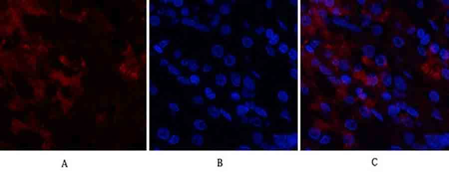
Fig.2. Immunofluorescence analysis of human stomach tissue. 1, Cdk4 Polyclonal Antibody (red) was diluted at 1:200 (4°C, overnight). 2, Cy3 Labeled secondary antibody was diluted at 1:300 (room temperature, 50min). 3, Picture B: DAPI (blue) 10min. Picture A: Target. Picture B: DAPI. Picture C: merge of A+B.

Fig.3. Immunofluorescence analysis of rat lung tissue. 1, Cdk4 Polyclonal Antibody (red) was diluted at 1:200 (4°C, overnight). 2, Cy3 Labeled secondary antibody was diluted at 1:300 (room temperature, 50min). 3, Picture B: DAPI (blue) 10min. Picture A: Target. Picture B: DAPI. Picture C: merge of A+B.

Fig.4. Immunohistochemical analysis of paraffin-embedded human brain, antibody was diluted at 1:100.

Fig.5. Immunohistochemical analysis of paraffin-embedded rat muscle, antibody was diluted at 1:100.
You must be logged in to post a review.


Reviews
There are no reviews yet.