| Product name | c-Fos Polyclonal Antibody |
| Immunogen | Synthesized peptide derived from human c-Fos around the non-phosphorylation site of S374 |
| Host | Rabbit |
| Reactivity | Human,Mouse,Rat |
| Applications | IF,WB,IHC,ELISA |
| Applications notes | Optimal working dilutions should be determined experimentally by the investigator. Suggested starting dilutions are as follows: IF 1:50-200;WB 1:500-1:2000;IHC 1:100-1:300;ELISA 1:5000;Not yet tested in other applications; |
| Clonality | Polyclonal |
| Preparation method | The antibody was affinity-purified from rabbit antiserum by affinity-chromatography using epitope-specific immunogen. |
| Alternative | FOS; G0S7; Proto-oncogene c-Fos; Cellular oncogene fos; G0/G1 switch regulatory protein 7 |
| Formulation | Liquid solution |
| Concentration | 1 mg/ml |
| Molecular weight | 62kD |
| Storage buffer | Liquid in PBS containing 50% glycerol, 0.5% BSA and 0.02% sodium azide. |
| Storage instructions | Stable for one year at -20°C from date of shipment. For maximum recovery of product, centrifuge the original vial after thawing and prior to removing the cap. Aliquot to avoid repeated freezing and thawing. |
| Shipping | Gel pack with blue ice. |
| Precautions | The product listed herein is for research use only and is not intended for use in human or clinical diagnosis. Suggested applications of our products are not recommendations to use our products in violation of any patent or as a license. We cannot be responsible for patent infringements or other violations that may occur with the use of this product. |
| Background | The Fos gene family consists of 4 members: FOS, FOSB, FOSL1, and FOSL2. These genes encode leucine zipper proteins that can dimerize with proteins of the JUN family, thereby forming the transcription factor complex AP-1. As such, the FOS proteins have been implicated as regulators of cell proliferation, differentiation, and transformation. In some cases, expression of the FOS gene has also been associated with apoptotic cell death. |
| Gene ID | 2353 |
| Alternative | FOS; G0S7; Proto-oncogene c-Fos; Cellular oncogene fos; G0/G1 switch regulatory protein 7 |
| Others | c-Fos Polyclonal Antibody detects endogenous levels of c-Fos protein. |
| Accession | P01100 |
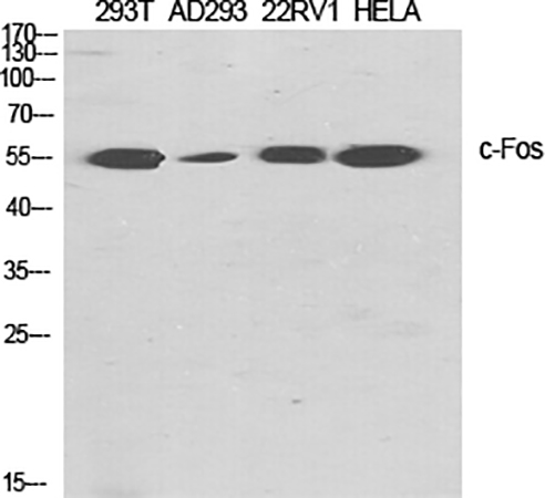
Fig.1. Western Blot analysis of 293T (1), AD293 (2), 22RV1 (3), Hela (4), diluted at 1:2000.
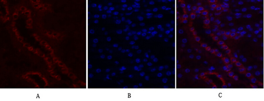
Fig.2. Immunofluorescence analysis of mouse kidney tissue. 1, c-Fos Polyclonal Antibody (red) was diluted at 1:200 (4°C, overnight). 2, Cy3 labeled secondary antibody was diluted at 1:300 (room temperature, 50min). 3, Picture B: DAPI (blue) 10min. Picture A: Target. Picture B: DAPI. Picture C: merge of A+B.
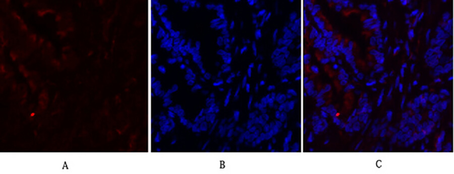
Fig.3. Immunofluorescence analysis of rat lung tissue. 1, c-Fos Polyclonal Antibody (red) was diluted at 1:200 (4°C, overnight). 2, Cy3 labeled secondary antibody was diluted at 1:300 (room temperature, 50min). 3, Picture B: DAPI (blue) 10min. Picture A: Target. Picture B: DAPI. Picture C: merge of A+B.
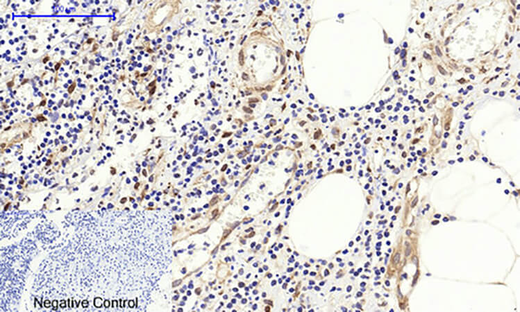
Fig.4. Immunohistochemical analysis of paraffin-embedded human appendix tissue. 1, c-Fos Polyclonal Antibody was diluted at 1:200 (4°C, overnight). 2, Sodium citrate pH 6.0 was used for antibody retrieval (>98°C, 20min). 3, secondary antibody was diluted at 1:200 (room temperature, 30min). Negative control was used by secondary antibody only.
You must be logged in to post a review.


Reviews
There are no reviews yet.