| Product name | Anti-Myc Tag Mouse Monoclonal Antibody (2D5) |
| Immunogen | Synthetic Peptide |
| Host | Mouse |
| Reactivity | Mammals, Bacteria |
| Applications | IF, IP, WB |
| Applications notes | Optimal working dilutions should be determined experimentally by the investigator. Suggested starting dilutions are as follows: WB (1:5000), IF (1:1000), IP (1:200). |
| Clonality | Monoclonal |
| Preparation method | The antibody was affinity-purified from mouse ascites by affinity-chromatography using specific immunogen |
| Alternative | Myc tag; Myc Epitope; c-Myc; bHLHe39; myc proto-oncogene protein; oncogene c-Myc |
| Formulation | Liquid solution |
| Storage buffer | Liquid in PBS, pH 7.4, containing 0.02% Sodium Azide as preservative and 50% Glycerol. |
| Storage instructions | Stable for one year at -20°C from date of shipment. For maximum recovery of product, centrifuge the original vial after thawing and prior to removing the cap. Aliquot to avoid repeated freezing and thawing. |
| Shipping | Gel pack with blue ice. |
| Precautions | The product listed herein is for research use only and is not intended for use in human or clinical diagnosis. Suggested applications of our products are not recommendations to use our products in violation of any patent or as a license. We cannot be responsible for patent infringements or other violations that may occur with the use of this product. |
| Background | Myc tag is a polypeptide protein tag derived from the c-myc gene product that can be added to a protein using recombinant DNA technology. It can be used for affinity chromatography, and then used to separate recombinant, overexpressed protein from wild type protein expressed by the host organism. It can also be used in the isolation of protein complexes with multiple subunits. |
| Alternative | Myc tag; Myc Epitope; c-Myc; bHLHe39; myc proto-oncogene protein; oncogene c-Myc |
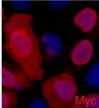
Fig.1. Immunofluorescence staining (1:2000) of Myc fusion protein in 293 cells with red and counterstained with DAPI.
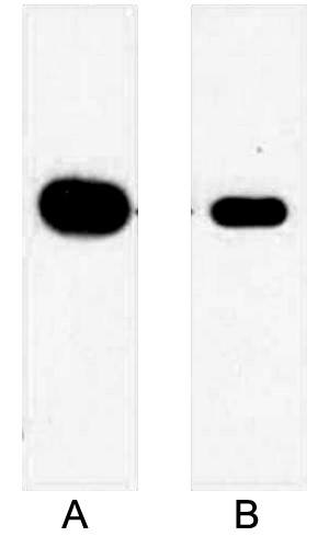
Fig.2. Western blot analysis of 1ug Myc fusion protein with Anti-Myc Mouse Monoclonal Antibody (2D5) in 1:5000 dilution (lane A) and 1:10000 dilution (lane B).
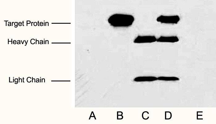
Fig.3. IP (1:200)-WB (1:5000) analysis of Myc fusion protein expression in 293 cells. Untransfected 293 cell lysate (lane A), transfected 293 cell lysate with Myc-tag protein (lane B); IP transfected 293 cell lysate with normal Mouse IgG (lane C) or with Anti Myc tag mAb (lane D) , and without both normal Mouse IgG and Myc tag mAb (lane E).
Author:Guo, Jinliang, et al. Publication name:Nature Communications IF:15
Author:Wang, Yifan, et al. Publication name:Advanced Science IF:14
Author:Cui Jie Publication name:International Journal of Biological Sciences IF:7.7
Author:黄宝玉 Publication name:Plant Biotechnol J IF:4
Author:Chen, Meng-Yan, et al. Publication name:Reproduction IF:3.7
Author:Yan, Qigui, et al. Publication name:Veterinary Sciences IF:2.3
You must be logged in to post a review.











Reviews
There are no reviews yet.