| Product name | Anti-Lamin B1 Monoclonal Antibody (15T1) |
| Immunogen | Recombinant Protein |
| Host | Mouse |
| Reactivity | Human, Mouse, Rat |
| Applications | IF, IHC, IP, WB |
| Applications notes | Optimal working dilutions should be determined experimentally by the investigator. Suggested starting dilutions are as follows: WB (1:2000-1:5000), IHC-P (1:50-1:300), IF (1:200), IP (1:200). |
| Clonality | Monoclonal |
| Preparation method | The antibody was affinity-purified from mouse ascites by affinity-chromatography using specific immunogen |
| Alternative | LMNB1; LMN2; LMNB; Lamin-B1 |
| Formulation | Liquid solution |
| Storage buffer | Liquid in PBS, pH 7.4, containing 0.02% Sodium Azide as preservative and 50% Glycerol. |
| Storage instructions | Stable for one year at -20°C from date of shipment. For maximum recovery of product, centrifuge the original vial after thawing and prior to removing the cap. Aliquot to avoid repeated freezing and thawing. |
| Shipping | Gel pack with blue ice. |
| Precautions | The product listed herein is for research use only and is not intended for use in human or clinical diagnosis. Suggested applications of our products are not recommendations to use our products in violation of any patent or as a license. We cannot be responsible for patent infringements or other violations that may occur with the use of this product. |
| Background | LMNB1 encodes one of the two B-type lamin proteins and is a component of the nuclear lamina. A duplication of LMNB1 is associated with autosomal dominant adult-onset leukodystrophy (ADLD). Alternative splicing results in multiple transcript variants. |
| Gene ID | 4001 |
| Alternative | LMNB1; LMN2; LMNB; Lamin-B1 |
| Accession | P20700 |
| Observed Band(KD) | 68 |
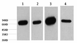
Fig.1. Western blot analysis of 1) HepG2, 2) 293T, 3) mouse brain tissue, 4) rat brain tissue, diluted at 1:5000.
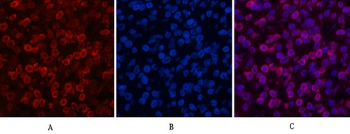
Fig.2 Immunofluorescence analysis of human lung cancer tissue. 1, Lamin B1 Monoclonal Antibody (15T1) (red) was diluted at 1:200 (4°C, overnight). 2, Cy3 Labeled secondary antibody was diluted at 1:300 (room temperature, 50min). 3, Picture B: DAPI (blue) 10min. Picture A: Target. Picture B: DAPI. Picture C: merge of A+B.
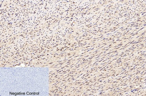
Fig.3. Immunohistochemical analysis of paraffin-embedded human uterus tissue. 1, Lamin B1 Monoclonal Antibody (15T1) was diluted at 1:200 (4°C, overnight). 2, Sodium citrate pH 6.0 was used for antibody retrieval (>98°C, 20min). 3, secondary antibody was diluted at 1:200 (room temperature, 30min). Negative control was used by secondary antibody only.
You must be logged in to post a review.


Reviews
There are no reviews yet.