| Product name | Anti-GAPDH Mouse Monoclonal Antibody (2B5) |
| Immunogen | Synthetic Peptide |
| Host | Mouse |
| Reactivity | Chicken, Dog, Hamster, Human, Monkey, Mouse, Pig, Rabbit, Rat, Sheep, Yeast |
| Applications | IF, IHC-P, WB |
| Applications notes | Optimal working dilutions should be determined experimentally by the investigator. Suggested starting dilutions are as follows: WB(1:10000-1:100000), IHC-P (1:400), IF (1:400). |
| Clonality | Monoclonal |
| Preparation method | The antibody was affinity-purified from mouse ascites by affinity-chromatography using specific immunogen |
| Alternative | GAPDH; GAPD; CDABP0047; OK/SW-cl.12; Glyceraldehyde-3-phosphate dehydrogenase; GAPDH; Peptidyl-cysteine S-nitrosylase GAPDH |
| Formulation | Liquid solution |
| Concentration | 1 mg/ml |
| Storage buffer | Liquid in PBS, pH 7.4, containing 0.02% Sodium Azide as preservative and 50% Glycerol. |
| Storage instructions | Stable for one year at -20°C from date of shipment. For maximum recovery of product, centrifuge the original vial after thawing and prior to removing the cap. Aliquot to avoid repeated freezing and thawing. |
| Shipping | Gel pack with blue ice. |
| Precautions | The product listed herein is for research use only and is not intended for use in human or clinical diagnosis. Suggested applications of our products are not recommendations to use our products in violation of any patent or as a license. We cannot be responsible for patent infringements or other violations that may occur with the use of this product. |
| Background | Glyceraldehyde 3-phosphate dehydrogenase (abbreviated as GAPDH or less commonly as G3PDH) is an enzyme of 37kDa that catalyzes the sixth step of glycolysis and thus serves to break down glucose for energy and carbon molecules. In addition to this long established metabolic function, GAPDH has recently been implicated in several non-metabolic processes, including transcription activation, initiation of apoptosis ER to Golgi vesicle shuttling, and fast axonal, or axoplasmic transport. |
| Gene ID | 2597 |
| Alternative | GAPDH; GAPD; CDABP0047; OK/SW-cl.12; Glyceraldehyde-3-phosphate dehydrogenase; GAPDH; Peptidyl-cysteine S-nitrosylase GAPDH |
| Accession | P04406 |
| Observed Band(KD) | 37 |
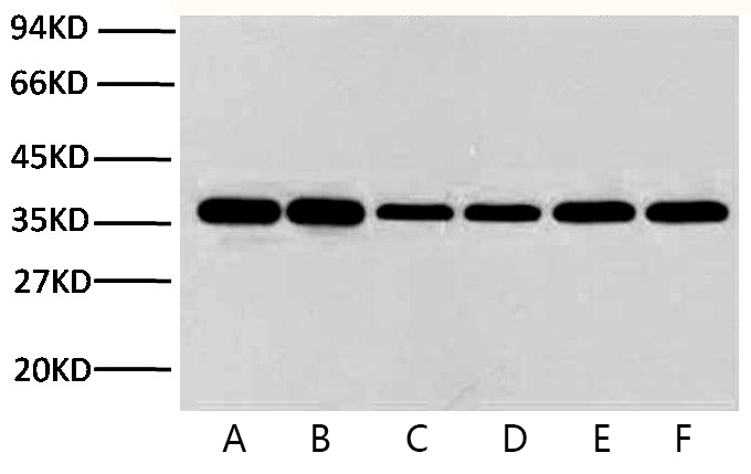
Fig.1. Western blot analysis of Hela (1), rat brain (2), rabbit muscle(3), sheep muscle(4), and mouse brain (5), diluted at 1:10000.
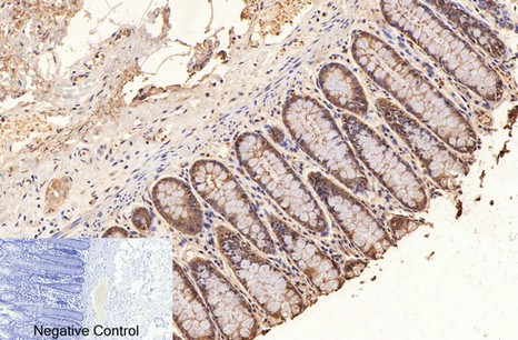
Fig.2. Immunohistochemical analysis of paraffin-embedded human colon tissue. 1, GAPDH Monoclonal Antibody (2B5) was diluted at 1:400 (4°C, overnight). Negative control was used by secondary antibody only.
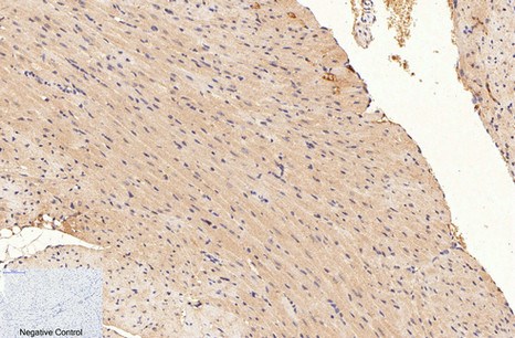
Fig.3. Immunohistochemical analysis of paraffin-embedded mouse heart tissue. 1, GAPDH Monoclonal Antibody (2B5) was diluted at 1:400 (4°C, overnight). Negative control was used by secondary antibody only.
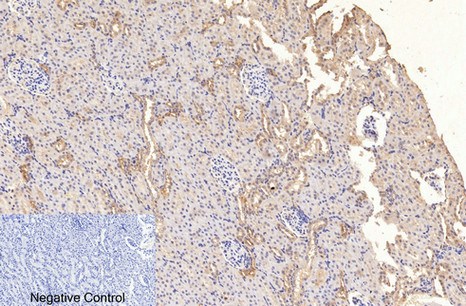
Fig.4. Immunohistochemical analysis of paraffin-embedded rat kidney tissue. 1, GAPDH Monoclonal Antibody (2B5) was diluted at 1:400 (4°C, overnight). Negative control was used by secondary antibody only.
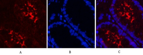
Fig.5. Immunofluorescence analysis of human colon tissue. 1, GAPDH Monoclonal Antibody (2B5) (red) was diluted at 1:400 (4°C, overnight). Picture A: Target. Picture B: DAPI. Picture C: merge of A+B.
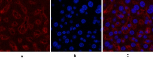
Fig.6. Immunofluorescence analysis of mouse liver tissue. 1, GAPDH Monoclonal Antibody (2B5) (red) was diluted at 1:400 (4°C, overnight). Picture A: Target. Picture B: DAPI. Picture C: merge of A+B.
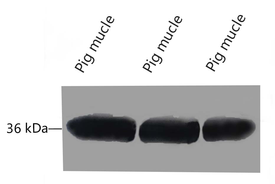
Fig.7 Western blot analysis of Pig muscle tissue, GAPDH Monoclonal Antibody (2B5) was diluted at 1:10000 (25°C, 3h).
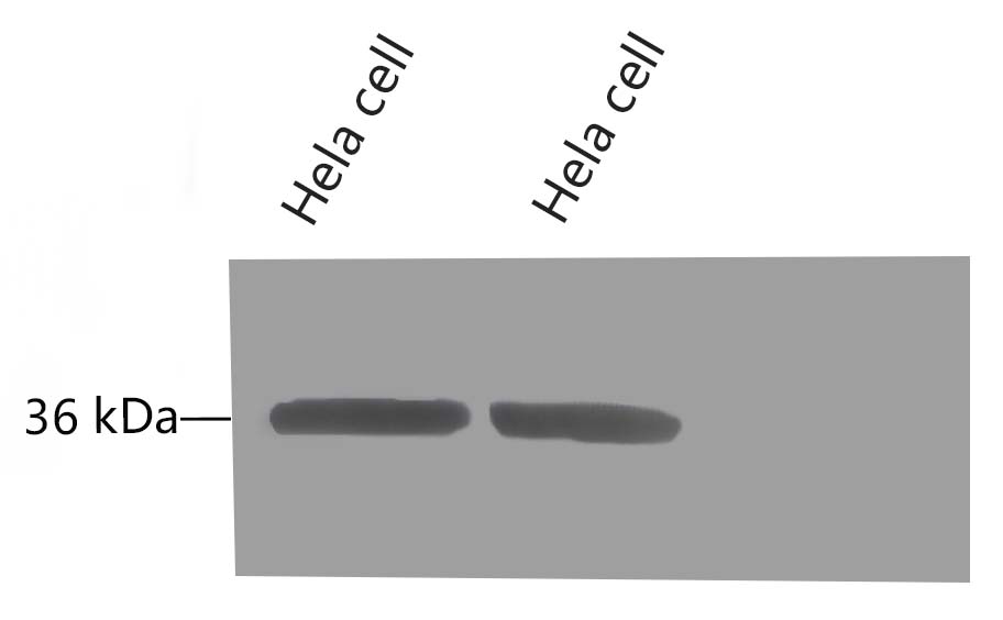
Fig.8 Western blot analysis of Hela, GAPDH Monoclonal Antibody (2B5) was diluted at 1:10000 (25°C, 3h).
Author:Wang, Xiafeng, et al. Publication name:Nature IF:48.5
Author:Yang X, Liu X J, Li Y Y, et al Publication name:Materials Science and Engineering: C IF:27
Author:Park, Suhyeon, et al. Publication name:Experimental & molecular medicine IF:13
Author:M Kim, S Park, S Kim, J Seo, S Roh Publication name:Am J Transl Res 2019;11(3):1555-1568 IF:12.9
Author:Qu, Zhihao, et al. Publication name:Food Chemistry IF:9.8
Author:Kim, Minseo, et al. Publication name:Biomaterials Research IF:9.6
Author:L Tian, P Tang, J Liu, Y Liu, L Hou, J Zhao, ... Publication name:Infect Genet Evol IF:8.2
Author:Z Qu, P Tian, L Wang, X Jin, M Guo, J Lu, ... Publication name:Sensors and Actuators B: Chemical IF:6.2
Author:Y Yang, R Gao, Z Zhu, W Xiao, J Wang, W Zhao, ... Publication name:International Immunopharmacology IF:6.1
Author:Liu X J, Yang B, Huang SN, et al Publication name:PLoS Pathog IF:6
Author:Xiong XX, Pan F, Chen RQ, et al Publication name:Cell Death Dis IF:6
Author:Yu L M, Li Z, Dong X, et al Publication name:Oxidative Medicine and Cellular Longevity IF:5
Author:Sui, Xiaolu, et al. Publication name:Mediators of Inflammation IF:5
Author:Niu, Xuan, et al Publication name:Arteriosclerosis, Thrombosis, and Vascular Biology (2017): ATVBAHA-116 IF:5
Author:Song F, Sun H, Huang L, et al Publication name:Cellular Physiology and Biochemistry IF:5
Author:X Gao, QP Li, JR Hao, K Sun, H Feng, ... Publication name:Sci Rep IF:5
Author:J Shi, C Li, Q Liang, Y Yao, Z Ji, M Zhou, J Cai, ... Publication name:Neural Regeneration Research IF:4.9
Author:J Xing, R Tan, F Huang, N Tian Publication name:J Comp Neurol IF:4.2
Author:Dong Z, Chen J, Yang X, et al Publication name:Oncotarget IF:4
Author:Liu F L, Mo E P, Yang L, et al Publication name:Oncotarget IF:4
Author:Chen, B, et al Publication name:Frontiers in Cellular Neuroscience 10(2016) IF:4
Author:Yin T, Zhang Z, Cao B, et al Publication name:Oncotarget IF:4
Author:Bai X L, Yang X Y, Li J Y, et al Publication name:2017, Oncotarget IF:4
Author:Yangfeng Chen, Zhijun Wang Publication name:Genes (Basel) IF:4
Author:Yu, Lin, et al Publication name:Applied and environmental microbiology 81 IF:4
Author:Huang, Hai-Jian, et al Publication name:Scientific reports 5 (2015) IF:4
Author:Shi Y, He M Publication name:Scientific reports IF:4
Author:Zheng, Liming, et al Publication name:Scientific Reports 6 (2016) IF:4
Author:Xia Zhong, Qian-Qian Wang, et al Publication name:Scientific Reports IF:4
Author:Peng H, Gong P G, Li J B, et al Publication name:Journal of translational medicine IF:4
Author:Zheng, T, et al Publication name:Journal of Ethnopharmacology 193(2016): 691-699 IF:4
Author:H Li, L Lv, S Tang, Y Zang, T Wan, D Wang, ... Publication name:Aquaculture IF:3.9
Author:Hu, Zhizhi, et al. Publication name:Renal Failure IF:3
Author:Huang, HJ, et al Publication name:Journal of Insect Physiology 98(2017):223-230 IF:3
Author:Min Xiang, Lei Zhang, et al Publication name:Gene IF:3
Author:Yihui Li, Hongdan Yan, et al Publication name:Biochemical and Biophysical Research Communications IF:3
Author:Zeng T, Zhou J, He L, et al Publication name:PloS one IF:3
Author:Chen W, Wang D, Du X, et al Publication name:Medical oncology IF:3
Author:Gao Q, Liu Y, Wu Y, et al Publication name:International journal of molecular medicine IF:3
Author:Wang C, Zeng N, Liu S, et al Publication name:PLoS One IF:3
Author:Han Y, Liu G, Jiang X, et al Publication name:Vaccine IF:3
Author:Liu W, Wang H, Wang Y, et al Publication name:Psychiatry research IF:3
Author:Li, Nan, et al Publication name:Experimental Cell Research 350 IF:3
Author:Zhang, Ke, et al. Publication name:Biochemistry and Biophysics Reports IF:3
Author:Wang M, Pan L, Zhou P, et al Publication name:PloS one IF:3
Author:Wu B, Wang S, Qin G, et al Publication name:J Mol Neurosci IF:3
Author:Binbin Yin, Zhenping Liu, et al Publication name:Oncology Reports IF:3
Author:Pan X, Mou J, Liu S, et al Publication name:Oncology reports IF:3
Author:Wang X, Yang L, Wu Y, et al Publication name:Environmental Toxicology and Chemistry IF:3
Author:Yang X, Chu H, Tang Y, et al Publication name:Neuroscience IF:3
Author:Zhang L, Chen P, Yang S, et al Publication name:Oncology Letters IF:2
Author:Fang K, Dong H, Jiang S, et al Publication name:Evidence-Based Complementary and Alternative Medicine IF:2
Author:Chen S, Li S, Chen L Publication name:Journal of medical virology IF:2
Author:Zhou Y F, Guo B, Ye M J, et al Publication name:Current eye research IF:2
Author:Zheng X, Li X, Lyu Y, et al Publication name:Medical Science Monitor: International Medical Journal of Experimental and Clinical Research IF:2
Author:Liu H, Li M, Cai S, et al Publication name:Acta Biochimica et Biophysica Sinica IF:2
Author:Wang, Yao, et al Publication name:Food Analytical Methods (2017): 1-7 IF:2
Author:X Tan, R Liu, D Zhao, Z He, W Li, M Zheng Publication name:Journal of advanced research IF:1
Author:Wang Y, Su K, Hu P C, et al Publication name:Ultrastructural pathology IF:1
Author:Y Liu, M Guo, Y Li, T Wang, Y Ren, R Wang, X Jiang Publication name:researchsquare IF:/
Author:VK Verma, S Malik, AK Sahu, V Prajapati, J Bhatia Publication name:researchsquare IF:0
Author:Zheng X T, Xiao X Q Publication name:The international journal of biochemistry & cell biology IF:/
Author:Zuo Y, Xiong N, Zhao H Publication name:Journal of Huazhong University of Science and Technology [Medical Sciences] IF:0
Author:Chen, Mengdi, et al. Publication name:npj Parkinson's Disease IF:
Author:Zhou, Heng, et al. Publication name:Biochimica et Biophysica Acta (BBA)-Molecular Basis of Disease IF:
Author:Shi X Z, Shi L J, Zhao Y R, et al Publication name:Developmental & Comparative Immunology IF:/
You must be logged in to post a review.


Reviews
There are no reviews yet.