| Product name | Anti-β-Actin Rabbit Polyclonal Antibody |
| Immunogen | Recombinant Protein |
| Host | Rabbit |
| Reactivity | Chicken, Human, Monkey, Mouse, Rabbit, Rat, Sheep, Xenopus laevis |
| Applications | IHC-P, WB |
| Applications notes | Optimal working dilutions should be determined experimentally by the investigator. Suggested starting dilutions are as follows: WB (1:1000), IHC-P (1:200). |
| Clonality | Polyclonal |
| Preparation method | The antibody was affinity-purified from rabbit antiserum by affinity-chromatography using specific immunogen |
| Alternative | ACTB; Actin, cytoplasmic 1; Beta-actin |
| Formulation | Liquid solution |
| Concentration | 1 mg/ml |
| Storage buffer | Liquid in PBS, pH 7.4, containing 0.02% Sodium Azide as preservative and 50% Glycerol. |
| Storage instructions | Stable for one year at -20°C from date of shipment. For maximum recovery of product, centrifuge the original vial after thawing and prior to removing the cap. Aliquot to avoid repeated freezing and thawing. |
| Shipping | Gel pack with blue ice. |
| Precautions | The product listed herein is for research use only and is not intended for use in human or clinical diagnosis. Suggested applications of our products are not recommendations to use our products in violation of any patent or as a license. We cannot be responsible for patent infringements or other violations that may occur with the use of this product. |
| Background | ACTB encodes one of six different actin proteins. Actins are highly conserved proteins that are involved in cell motility, structure, and integrity. This actin is a major constituent of the contractile apparatus and one of the two nonmuscle cytoskeletal actins. |
| Gene ID | 60 |
| Alternative | ACTB; Actin, cytoplasmic 1; Beta-actin |
| Others | The antibody detects endogenous β-actin protein. |
| Accession | P60709 |
| Observed Band(KD) | 43 |
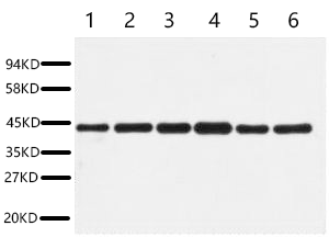
Fig.1. Western blot analysis of HepG2 (1), Rat liver (2), Mouse kidney (3), Rabbit testic (4), Sheep lung (5), 293T (6), diluted at 1:5000. Secondary antibody was diluted at 1:20000.
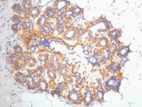
Fig.2. IHC-P staining of Human ovary tissue, diluted at 1:200.
Author:Duan, Shiwen, et al. Publication name: Nano Today IF:10.9
Author:Zhang N, Yin Y, Liu X, et al Publication name:Plant Physiol IF:6
Author:Zhao, Xinyu, et al. Publication name:Colloids and Surfaces B: Biointerfaces IF:5
Author:Y Tang, Y Liu, H Zhou, H Lu, Y Zhang, J Hua, ... Publication name:International Journal of Molecular and Cellular Medicine IF:4.2
Author:He F F, Bao D, Su H, et al Publication name:J Cell Physiol IF:4
Author:DM Xu, S He, XF Liang, JQ Wu, ... Publication name:The EMBO Journal IF:4
Author:Yang, Dongxue, et al Publication name:Frontiers in Molecular Neuroscience 9 (2016) IF:4
Author:Zuo Y C, Xiong N X, Shen J Y, et al Publication name:Neurochemical Research IF:3
Author:Li H, Chen L, Wang T, et al Publication name:PLoS One IF:3
Author:Wang J H, Zhou W W, Cheng S T, et al Publication name:Molecular medicine reports IF:2
Author:Ben-Xiang Qi, Ying-Jian Chen, et al Publication name:Cytotechnology IF:2
You must be logged in to post a review.


Reviews
There are no reviews yet.