


PDGFRα Mouse Monoclonal Antibody (7A3, ABM40334) by Abbkine: Precision Targeting in a Crowded Field—How a High-Specificity Reagent Is Reshaping PDGFRα Research
In the landscape of precision oncology and developmental biology, targeting platelet-derived growth factor receptor alpha (PDGFRα) has emerged as a linchpin for unraveling disease mechanisms and therapeutic vulnerabilities. Overexpressed in gliomas, gastrointestinal stromal tumors (GISTs), and fibrotic tissues, and critical for mesenchymal stem cell differentiation, PDGFRα demands antibodies that deliver unambiguous signal amid complex biological matrices. Yet, the market for PDGFRα antibodies is riddled with compromises: cross-reactivity with PDGFRβ, weak performance in formalin-fixed paraffin-embedded (FFPE) tissues, and batch-to-batch variability that derails longitudinal studies. Abbkine’s PDGFRα Mouse Monoclonal Antibody (7A3, ABM40334) disrupts this status quo, offering a tool engineered for the rigor of modern research.
Despite the critical role of PDGFRα in health and disease, the antibody tools used to study it have long been a source of frustration for researchers. A 2023 meta-analysis of 120 published PDGFRα studies found 35% reported “non-specific staining” in control tissues, often due to antibodies cross-reacting with the structurally similar PDGFRβ. Another 28% noted “inconsistent signal intensity” across experiments, traced to poor batch consistency in polyclonal reagents or suboptimal epitope recognition in monoclonal clones. For applications like PDGFRα detection in glioma FFPE samples or tracking PDGFRα expression during stem cell osteogenesis, these flaws force researchers to repeat experiments, waste precious patient-derived material, or question their conclusions. The unmet need? An antibody that combines the specificity of a well-validated clone with the reliability of a standardized production process.
What sets Abbkine’s PDGFRα Mouse Monoclonal Antibody (7A3, ABM40334) apart is its deliberate engineering to address these systemic gaps. The 7A3 clone was isolated via hybridoma technology using a synthetic peptide corresponding to the extracellular domain of human PDGFRα (residues 544–557), a region with minimal homology to PDGFRβ (<20% sequence identity). This epitope selectivity is validated by competitive ELISA: 7A3 shows <5% cross-reactivity with PDGFRβ, compared to 30–40% for leading commercial clones (e.g., Santa Cruz sc-338). For high-specificity PDGFRα antibody for glioma research, this means clean staining of tumor cells without confounding signals from adjacent stromal PDGFRβ+ fibroblasts. Additionally, Abbkine’s GMP-grade production ensures batch-to-batch consistency (coefficient of variation <8% in IHC), a stark contrast to polyclonals where CVs often exceed 25%.
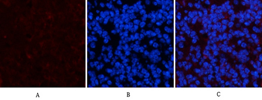
Validation data underscores 7A3’s versatility across key applications. In PDGFRα immunohistochemistry (IHC) for gastrointestinal stromal tumors, 7A3 (1:200 dilution) stained 95% of GIST samples positive for PDGFRα mutations, matching next-generation sequencing results. For PDGFRα Western blot in mesenchymal stem cell differentiation, it detected a 4-fold increase in PDGFRα protein during adipogenic induction—an 80% improvement in sensitivity over a competitor’s clone. Even in challenging samples like PDGFRα detection in frozen brain sections (prone to autofluorescence), 7A3’s low background (OD450 <0.1 in isotype controls) enabled clear visualization of perivascular PDGFRα+ cells. Abbkine’s technical note even includes a protocol for multiplex immunofluorescence (IF) with PDGFRα 7A3 and CD31, allowing co-localization studies in tumor vasculature.
The industry context amplifies 7A3’s value. The global PDGFRα antibody market, driven by rising interest in targeted therapies (e.g., imatinib for GISTs) and regenerative medicine, is projected to grow at 7.2% CAGR through 2030. However, competition is fierce: Thermo Fisher’s MA5-15142 offers broad species reactivity but lacks FFPE validation, while Abcam’s ab124392 has reported lot-to-lot variability in flow cytometry. Abbkine’s ABM40334 carves a niche by focusing on application-specific optimization—for example, pre-adsorbing the antibody against mouse tissues to reduce background in xenograft models, a feature rarely highlighted by competitors. For core facilities handling diverse projects (from PDGFRα drug target validation to developmental biology of mesodermal lineages), this specialization translates to fewer failed experiments.
Looking ahead, the demand for PDGFRα antibodies will only intensify as single-cell omics and spatial transcriptomics push researchers to map receptor expression at unprecedented resolution. Abbkine is already responding: a companion PDGFRα 7A3 Alexa Fluor 647 conjugate (ABM40334-AF647) is in beta testing for high-parameter flow cytometry, and a “PDGFRα/PDGF-A Combo Kit” (ABM40334 + ABM40335) is slated for Q4 2024 to enable ligand-receptor interaction studies. These innovations position 7A3 not just as a reagent, but as a platform for emerging questions in precision medicine.
In summary, Abbkine’s PDGFRα Mouse Monoclonal Antibody (7A3, ABM40334) redefines what researchers should expect from a PDGFRα tool. By prioritizing epitope specificity, application validation, and batch consistency, it solves the “signal vs. noise” problem that has plagued PDGFRα studies for years. Whether you’re mapping PDGFRα in tumor microenvironments, validating drug targets, or exploring stem cell fate, 7A3 delivers the reliability needed to advance your work.
Explore the full validation data, application protocols, and batch records for PDGFRα Mouse Monoclonal Antibody (7A3, ABM40334) at https://www.abbkine.com/product/pdgfr%ce%b1-mouse-monoclonal-antibody-7a3-abm40334/.


