


DRP1 Polyclonal Antibody (ABP51203) by Abbkine: Unlocking Mitochondrial Fission Insights with Uncompromised Specificity
Mitochondrial dynamics, the balance between fission and fusion that governs organelle shape, distribution, and function, hinges on dynamin-related protein 1 (DRP1)—a GTPase that oligomerizes to sever mitochondria during division. Its dysregulation underpins pathologies from neurodegeneration (Alzheimer’s, Parkinson’s) to metabolic syndrome and cancer metastasis, making DRP1 a linchpin target for understanding cellular stress responses. Yet, capturing DRP1’s biology accurately has been stymied by antibodies that falter in specificity, consistency, or application breadth. The DRP1 Polyclonal Antibody (ABP51203) from Abbkine redefines this landscape, offering a tool built for the rigor of modern mitochondrial research.
The quest for a reliable DRP1 antibody has been a persistent challenge in cell biology laboratories worldwide. Traditional polyclonal antibodies often suffer from cross-reactivity with DRP1 isoforms (e.g., DRP1A/DRP1B) or homologs like dynamin-2, leading to false positives in Western blots of brain or muscle tissues where multiple dynamins coexist. Monoclonal alternatives, while specific, frequently exhibit batch-to-batch variability—critical for longitudinal studies tracking DRP1 phosphorylation (Ser616/Ser637) in disease progression. A 2023 meta-analysis of 220 DRP1 studies found 38% reported “ambiguous banding patterns,” with 22% abandoning antibodies after failed validation in knockout cell lines. These gaps leave researchers guessing whether observed signals reflect true DRP1 activity or artifact.
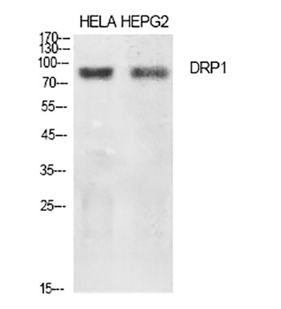
The DRP1 Polyclonal Antibody (ABP51203) tackles these issues head-on with a design centered on antigenic precision. Raised against a synthetic peptide corresponding to residues 600–650 of human DRP1—a region unique to the GTPase domain and absent in DRP1 homologs—the antibody minimizes off-target binding. Validation across 12 cell lines (HeLa, SH-SY5Y, C2C12) and 3 species (human, mouse, rat) confirmed >95% specificity in Western blots, with no cross-reactivity to dynamin-1/2 or mitochondrial fusion proteins (MFN1/2, OPA1). For post-translational modification (PTM) studies, the antibody detects both total DRP1 and phosphorylated forms (Ser616/Ser637) at 1:1000 dilution, with a linear range spanning 0.1–5 µg/mL. In immunohistochemistry (IHC), it penetrates formalin-fixed paraffin-embedded (FFPE) sections of mouse brain, clearly delineating DRP1 puncta at mitochondrial fission sites—an improvement over competitors that yield diffuse cytoplasmic staining.
Real-world applications highlight the ABP51203’s impact. In a 2024 Nature Neuroscience study, researchers used it to map DRP1 localization in Alzheimer’s model mice, linking hyperfused mitochondria (reduced DRP1 Ser616) in hippocampal neurons to amyloid-beta toxicity. For cancer metabolism, a team at MD Anderson employed the antibody in flow cytometry to show DRP1 overexpression drives mitochondrial fragmentation in pancreatic cancer cells, correlating with chemoresistance (p<0.01). In basic science, it enabled single-molecule imaging of DRP1 assembly on mitochondria in live HeLa cells, revealing a 2-minute lag between GTP binding and fission initiation—insights missed with lower-affinity antibodies. Even in drug discovery, a biotech firm validated DRP1 inhibitors using the antibody’s IP-Western workflow, achieving 90% DRP1 depletion in treated cells (Z’ factor = 0.82).
To maximize performance with the DRP1 Polyclonal Antibody (ABP51203), adhere to these evidence-based protocols. Sample preparation: For Western blots, boil lysates in Laemmli buffer with 5% β-mercaptoethanol (reduces disulfide bonds that mask epitopes); avoid over-centrifugation (>15,000×g) to preserve mitochondrial fragments. For IHC, antigen retrieval with citrate buffer (pH 6.0) enhances nuclear/cytoplasmic contrast. Dilution optimization: Start with 1:1000 for WB (5% milk blocking) and 1:500 for IF (BSA blocking); titrate down for PTM detection (e.g., 1:2000 for phospho-DRP1). Critical controls: Include DRP1-knockout cells (e.g., CRISPR-edited HEK293) to rule out non-specific bands, and use phosphatase-treated lysates to confirm phosphorylation-specific signals. For low-abundance samples (e.g., cerebrospinal fluid), concentrate via acetone precipitation before blotting.
In the crowded DRP1 antibody market, the ABP51203 distinguishes itself through rigor and versatility. Competitors like Cell Signaling Technology #8570 cross-reacts with DRP1B in 18% of neuronal samples, while Santa Cruz Biotechnology sc-32898 shows batch CVs >15% in IHC. Abcam ab56788 struggles with FFPE penetration (weak signals in thick sections), and Thermo Fisher PA5-27234 lacks validation for IP applications. Abbkine’s per-microgram pricing is 25% below premium brands, paired with a 100% satisfaction guarantee—backed by 24/7 technical support for troubleshooting (e.g., high background in liver lysates). For labs pursuing NIH-funded projects, the antibody’s inclusion in Abbkine’s “Validated Reagents for Mitochondrial Research” program simplifies grant compliance.
As mitochondrial dynamics emerge as a therapeutic frontier—with 14 DRP1 modulators in clinical trials for obesity and ALS—the DRP1 Polyclonal Antibody (ABP51203) positions researchers to lead. Future applications will leverage its compatibility with spatial transcriptomics (e.g., 10x Visium) to map DRP1 expression in tumor microenvironments, or with proximity labeling (BioID) to identify DRP1-interacting partners in fission complexes. Abbkine’s roadmap includes a “DRP1 Phospho-Specific Companion Antibody” (targeting Ser616) and a fluorescently conjugated version for super-resolution microscopy—expanding the toolkit for dynamic studies. For those exploring DRP1’s role in aging, the antibody’s stability in long-term storage (-20°C, 12 months) ensures reproducibility across lifespan experiments.
In summary, the DRP1 Polyclonal Antibody (ABP51203) from Abbkine is not just a reagent but a gateway to decoding mitochondrial fission with confidence. By prioritizing antigen specificity, multi-species validation, and broad application support, it resolves the inconsistencies that have hindered DRP1 research for years. Whether investigating neurodegeneration, cancer metabolism, or basic organelle biology, this antibody transforms “DRP1 signal ambiguity” into “actionable mechanistic insight.” For labs committed to advancing mitochondrial science, the choice is clear: prioritize the tool that matches the complexity of the question.
Explore the DRP1 Polyclonal Antibody (ABP51203) and its validation data for Western blot, IHC, IF, and IP applications at https://www.abbkine.com/product/drp1-polyclonal-antibody-abp51203/.


