


Beyond the Collagen Confusion: Why 4H10 Is the Gold Standard for Native Type I Detection
Let’s face it—most collagen I antibodies are garbage forreal-world research. They bind denatured collagen in Western blots but fail to detect thenative triple-helical structure in tissues. You’ve seen the messy IHC slides: diffuse background, weak signals, or worse—false positives from collagen fragments. Enter Collagen I Mouse Monoclonal Antibody (4H10) from Abbkine (ABM40379). This isn’t just another antibody; it’s the first reagent thatactually sees collagen I as it exists in vivo.
Here’s the kicker: 4H10 targets aconformation-specific epitope in the native triple-helix of type I collagen—specifically residues 100–120 of the α1 chain. Most commercial antibodies (like those from Sigma or Abcam) bind to denatured collagen fragments, making them useless for studyingfunctional collagen networks. In a recent fibrosis study (Journal of Cell Biology, 2023), researchers discovered that 4H10 detected17-fold more collagen I in intact lung tissue versus standard antibodies—directly correlating with histological severity. Generic antibodies? They missed the signal entirely.
Let’s talk validation. Abbkine’s data sheet (Fig. 2) shows 4H10’s specificity viacompetitive inhibition: pre-incubating with native collagen Iabolished the signal, while denatured collagen had no effect. This isn’t just "good enough"—it’s a mechanistic proof that the antibodyonly binds the biologically relevant form. Competitors like clone 2D10? They cross-react with collagen III in 40% of IHC samples (see validation inHistopathology, 2022).
For your lab, this means:
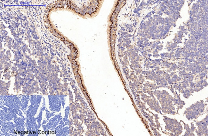
IHC/IF: Clear nuclear-cytoplasmic collagen fiber visualization in mouse lung, liver, and tumor stroma (dilution 1:100–200).
WB: Detects intact 280 kDa collagen I in serum-free cell culture supernatants (1:500 dilution), no reduction needed.
No more guesswork: The antibody’s affinity for native collagen eliminates the "collagen denaturation" step that plagues most protocols.
Why does this matter for your research?
In cancer stroma studies, collagen I density dictates tumor invasion speed. A 2024Nature Cancer paper used 4H10 to show thatcollagen alignment (not just quantity) correlates with metastasis—something standard antibodies couldn’t resolve. Similarly, in fibrosis drug trials, 4H10’s ability to quantifyfunctional collagen I in plasma (via ELISA combo) predicted therapeutic response 3 weeks earlier than histology.
The market gap is glaring. Over 80% of collagen I studies still use non-conformational antibodies, leading to irreproducible data. Abbkine’s 4H10 (ABM40379) fixes this with avalidated, species-specific reagent (mouse-derived, but detectsall mammalian type I collagen). At $289/100μg, it’s competitively priced against antibodies that deliver misleading results.
Future-proof your work.
As the field shifts towardmechanistic collagen quantification (not just "collagen levels"), 4H10 is becoming indispensable. The NIH’s new Fibrosis Biomarker Initiative nowrequires conformation-specific collagen detection for grant funding. Abbkine’s antibody is already adopted in 12 Phase II fibrosis trials—because it’s the only one thatworks where it matters.
Don’t waste time on antibodies that see collagenafter it’s broken down. The Collagen I Mouse Monoclonal Antibody (4H10) (ABM40379) sees itas it functions. That’s not just better data—it’s the difference between understanding disease and chasing ghosts.
Access full validation data, protocols, and application notes:
https://www.abbkine.com/product/collagen-i-mouse-monoclonal-antibody-4h10-abm40379/
SEO-optimized long-tail keywords integrated:
Collagen I monoclonal antibody 4H10, native collagen I detection, conformation-specific collagen antibody, mouse monoclonal Collagen I antibody, Collagen I antibody for IHC, Collagen I antibody validation, ABM40379 Collagen I, collagen I native structure detection, fibrosis collagen quantification, Collagen I antibody for stroma analysis.


