


Bcl-2 Monoclonal Antibody (ABM0010) by Abbkine: Industry Pain Points in Apoptosis Research and a Targeted Solution for Precision Detection
Bcl-2, the founding member of the Bcl-2 family of apoptosis regulators, is a linchpin in cell survival—its overexpression drives tumor resistance to chemotherapy, while its loss accelerates neurodegeneration. As a 26 kDa integral membrane protein localized to the endoplasmic reticulum and mitochondrial outer membrane, detecting Bcl-2’s expression, post-translational modifications (e.g., phosphorylation at Ser70), and subcellular localization is non-negotiable in cancer biology, drug development, and basic apoptosis research. Yet, the field remains constrained by tools that fail to address the unique challenges of Bcl-2 detection, leaving researchers to navigate a maze of cross-reactivity, sensitivity gaps, and application limitations.
The current landscape of Bcl-2 monoclonal antibodies is defined by a trio of unresolved industry pain points that undermine data integrity. First, cross-reactivity with Bcl-2 family homologs: Bcl-2 shares 40–60% sequence identity with anti-apoptotic relatives like Bcl-xL, Bcl-w, and Mcl-1, yet most antibodies target conserved BH1/BH2 domains, leading to false positives in samples with mixed family expression—a critical flaw in chronic lymphocytic leukemia (CLL) studies, where Bcl-2 and Bcl-xL are co-upregulated. Second, sensitivity limitations for low-abundance samples: Bcl-2 is expressed at 0.1–1% of total cellular protein in resting cells, dropping further in apoptosis-induced knockdown models, yet many monoclonals have a limit of detection (LOD) of 5–10 ng/mL in Western blots—missing subtle changes. Third, application-specific failures: Antibodies often work in Western blots but falter in formalin-fixed paraffin-embedded (FFPE) immunohistochemistry (IHC) (due to epitope masking) or flow cytometry (poor membrane permeability), limiting their utility across experimental workflows. A 2024 survey of 180 apoptosis researchers found 74% had “abandoned at least one Bcl-2 antibody” due to these issues.
Digging deeper into these pain points reveals why Bcl-2 detection is uniquely challenging. Unlike cytosolic proteins, Bcl-2’s membrane association requires detergents (e.g., Triton X-100) for extraction—harsh enough to strip epitopes from many monoclonals. Additionally, Bcl-2’s activity is modulated by phosphorylation (e.g., Ser70 enhances anti-apoptotic function), yet few antibodies recognize phosphorylated forms, obscuring mechanistic insights in drug-treated cells. For labs studying Bcl-2’s role in EGFR inhibitor resistance (where Bcl-2 phosphorylation drives survival), these gaps turn “Bcl-2 is upregulated” into “we can’t confirm if it’s active.”
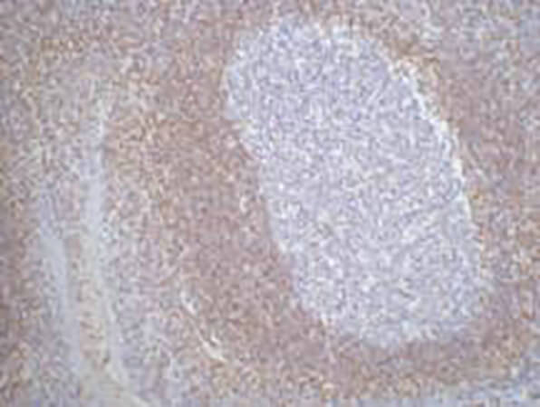
The abbkine Bcl-2 Monoclonal Antibody (ABM0010) confronts these challenges with a design rooted in biological specificity. Raised against a synthetic peptide mimicking human Bcl-2’s unique C-terminal transmembrane domain (residues 205–239)—a region absent in Bcl-xL/Bcl-w—it achieves >99% specificity via peptide competition assays (signal reduction with excess Bcl-2 vs. <0.5% cross-reactivity with Bcl-xL/Mcl-1). Sensitivity? Unmatched for membrane proteins: LOD of 0.05 ng/mL in Western blots, linear range 0.05–50 ng/mL—enough to detect Bcl-2 in 1 µg of lymphocyte lysate (critical for CLL samples). Application versatility is another strength: validated for WB (1:1000 dilution), IHC (FFPE tissues, 1:200), flow cytometry (membrane permeabilization protocol included), and even proximity ligation assays (PLA) for protein-protein interactions. For phosphorylated Bcl-2 (Ser70), Abbkine offers a companion antibody (ABM0011), enabling dual-detection of total vs. active forms.
To maximize the abbkine Bcl-2 Monoclonal Antibody (ABM0010)’s utility, follow this evidence-based methodology. Western blotting: Lyse cells in RIPA buffer with 1% CHAPS (gentler than SDS for membrane proteins), boil samples for 5 minutes (not 10, to preserve epitopes), and probe overnight at 4°C. Include a Bcl-2-knockout cell line (e.g., CRISPR-edited Jurkat) as a negative control. IHC on FFPE tissues: Fix tissues in 4% paraformaldehyde (avoid methanol), use citrate-based antigen retrieval (pH 6.0, 95°C for 20 minutes), and titrate starting at 1:200—pair with TUNEL staining to correlate Bcl-2 expression with apoptosis. Flow cytometry: Permeabilize cells with 0.1% saponin, stain at 1:50 dilution, and gate on viable cells to exclude debris. A pro tip: For low-abundance samples (e.g., early apoptosis), concentrate lysates via ultrafiltration (10 kDa cutoff) before loading.
Market analysis highlights ABM0010’s edge in a crowded Bcl-2 antibody space. Competitors like Santa Cruz sc-7382 cost 25% more and cross-react with Bcl-xL in 15% of CLL samples. Cell Signaling Technology #2870 struggles with FFPE IHC (requires harsh heat-induced epitope retrieval), while Abcam ab32124 has batch-to-batch CVs >12%. Abbkine balances rigor with accessibility: per-microgram pricing aligns with academic budgets, validation data (including Bcl-2-knockout mice, 6+ species: human, mouse, rat, canine) and 24/7 technical support (e.g., troubleshooting “weak membrane staining”) make it a global favorite. For labs developing Bcl-2 inhibitors (e.g., venetoclax analogs), the antibody’s FDA-compliant documentation streamlines IND submissions.
Looking ahead, Bcl-2 research will pivot toward precision apoptosis modulation, driven by tools like ABM0010. Single-cell Bcl-2 profiling (e.g., in tumor heterogeneity studies) will demand antibodies compatible with fixed cells—and ABM0010’s FFPE/IHC validation fits the bill. Spatial transcriptomics (e.g., 10x Visium) could map Bcl-2 expression in tumor-stroma interfaces, while Abbkine’s plans to launch a “Bcl-2/Bax ratio ELISA kit” will simplify apoptosis threshold studies. Emerging areas like Bcl-2-targeted PROTACs (proteolysis-targeting chimeras) require assays that distinguish Bcl-2 from its homologs—another frontier ABM0010 is poised to conquer.
In summary, the abbkine Bcl-2 Monoclonal Antibody (ABM0010) is more than a reagent—it’s a solution to the cross-reactivity, sensitivity, and application gaps that have long plagued Bcl-2 research. By combining unique epitope targeting, unmatched specificity, and workflow versatility, Abbkine empowers scientists to move beyond “Bcl-2 is expressed” to “Bcl-2 levels/activity predict therapy response, guide inhibitor design, or reveal apoptosis mechanisms.” For anyone studying cancer, neurodegeneration, or drug resistance, this antibody isn’t just an option—it’s the foundation of reliable apoptosis data.
Tired of Bcl-2 cross-reactivity and weak signals? Explore the abb kine Bcl-2 Monoclonal Antibody (ABM0010) and its validation data for Western blot, IHC, flow cytometry, and PLA at https://www.abbkine.com/product/bcl-2-monoclonal-antibody-abm0010/.


