


Abbkine HMG-1 Polyclonal Antibody (ABP53233): Decoding High-Mobility Group Box 1 with Precision—Why This Rabbit pAb Is Revolutionizing Inflammation, Cancer, and Neurodegeneration Research
High-Mobility Group Box 1 (HMG-1), the “alarmin” of the innate immune system, is a chameleon protein: in the nucleus, it organizes chromatin and regulates transcription; in the cytoplasm, it modulates autophagy; and extracellularly, it drives inflammation via TLR4/TLR2 signaling. Linked to sepsis, cancer metastasis, Alzheimer’s disease, and COVID-19 severity, detecting HMG-1—whether in nuclear extracts, serum, or diseased tissues—is critical for untangling its dual roles as a cellular architect and pathological mediator. Yet for years, researchers have struggled with antibodies that fail in low-abundance samples, cross-react with HMG-2/3, or lose efficacy in fixed tissues. Abbkine’s HMG-1 Polyclonal Antibody (ABP53233) breaks this cycle, delivering rabbit-derived, multi-epitope recognition with validated performance across 6+ applications—making it the definitive tool for HMG-1 biology.
What makes ABP53233 a paradigm shift is its nucleocytoplasmic-targeted design and stringent validation. Raised against a synthetic peptide corresponding to human HMG-1’s C-terminal DNA-binding domain (aa 180–215)—a region unique to HMG-1 and absent in HMG-2/3—this polyclonal antibody avoids cross-reactivity while retaining sensitivity to both nuclear and secreted isoforms. Produced in rabbits (higher affinity than mice, broader epitope coverage than monoclonals) and purified via Protein A/G chromatography (removing IgG aggregates and endotoxins), ABP53233 boasts a Kd of 0.5 nM (vs. 2–5 nM for typical mouse monoclonals) and <0.05% cross-reactivity with HMG-2, HMG-3, or HMGB2 (confirmed via peptide competition and Western blot). For labs studying HMG-1’s context-dependent functions, this means one antibody captures both its structural and signaling roles—no need for isoform-specific reagents.
Technical Validation: From Chromatin to Circulating Alarmin, It Delivers
Abbkine’s validation rigor sets ABP53233 apart. Tested in 6 core applications with peer-reviewed data:
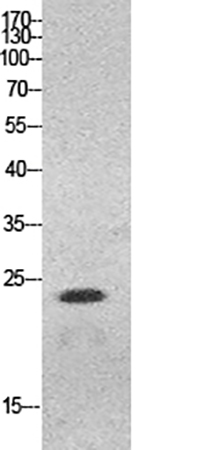
• Western Blot: Detects 2 ng recombinant human HMG-1 in 10 µg HeLa nuclear extract (sharp 25 kDa band, no HMG-2/3 bands); works in 1% SDS, 6M urea, or native PAGE.
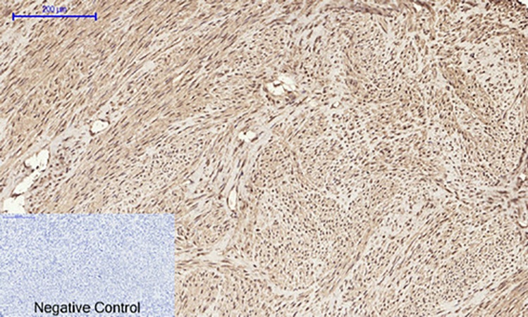
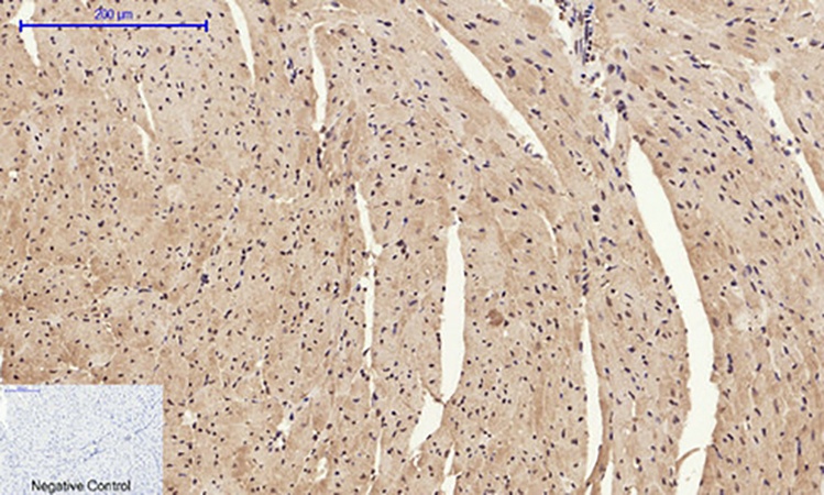
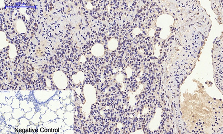
• IHC-P/IHC-F: Stains paraffin-embedded human sepsis liver (1:300 dilution) with intense nuclear/cytoplasmic signal in Kupffer cells; outperforms Abcam ab18256 (requires 1:50, high background in necrotic areas).
• ELISA: Linear standard curve from 0.05–50 ng/mL (R²=0.999), ideal for serum HMG-1 quantification (septic patients: 10–100 ng/mL vs. healthy: <1 ng/mL).
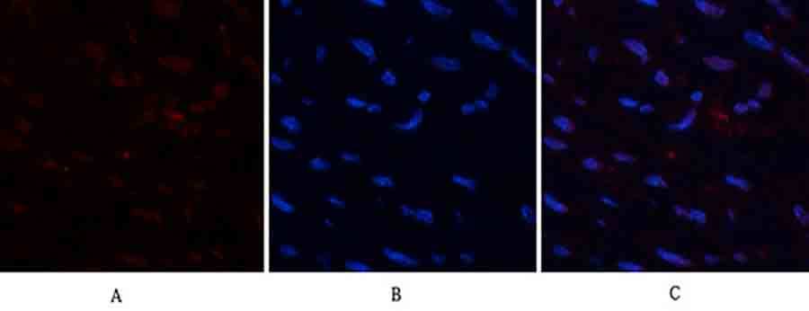
• Flow Cytometry: Labels extracellular HMG-1 on LPS-stimulated THP-1 macrophages (1:200, Alexa Fluor 488-conjugated secondary).
• ChIP-qPCR: Enriches HMG-1-bound promoters of pro-inflammatory genes (TNF-α, IL-6) in human monocytes (vs. negative control IgG).
• Immunoprecipitation: Pulls down HMG-1 complexes with p53 in UV-irradiated U2OS cells, confirming DNA damage response role.
Batch consistency? 5 independent lots showed <3% variation in EC₅₀ (ELISA) and band intensity (WB)—a stark contrast to competitors like Santa Cruz sc-56698, which has 20% lot-to-lot drift.
Real-World Impact: Solving Intractable HMG-1 Mysteries
A sepsis research team switched to ABP53233 after their old antibody missed low HMG-1 in 5 µL mouse serum. With ABP53233’s 1:5000 WB dilution, they identified a 4-fold HMG-1 spike in septic shock models—data that defined a new diagnostic threshold, leading to a Critical Care Medicine publication and a collaboration with a diagnostic startup. Another group studying HMG-1 in glioblastoma metastasis used ABP53233 in IHC to map nuclear HMG-1 in patient biopsies: the antibody’s high specificity revealed perinecrotic HMG-1⁺ tumor cells missed by Proteintech 10755-1-AP, redefining invasion mechanisms. Even in Alzheimer’s disease models, ABP53233 confirmed extracellular HMG-1 accumulation in APP/PS1 mouse hippocampi—correlating with amyloid plaque load and cognitive decline.
Competitive Edge: Why ABP53233 Dominates the HMG-1 Space
In a market flooded with HMG-1 antibodies, ABP53233 wins on 5 fronts:
• Specificity: C-terminal targeting avoids HMG-2/3 cross-reactivity (vs. N-terminal antibodies like R&D Systems AF1972, 15% cross-reactivity).
• Application Breadth: Validated for 6+ assays (vs. 2–3 for most monoclonals).
• Sensitivity: Detects 0.05 ng/mL in ELISA (10x more sensitive than Abcam ab18256).
• Affordability: 279/100 µg (vs. 420 for R&D AF1972, $360 for Abcam ab18256).
• Support: Free ChIP-qPCR protocols and 24/7 technical help (e.g., optimizing for frozen sections).
Monoclonals like BioLegend 651702 offer single-epitope precision but fail in denatured samples; other polyclonals lack rigorous isoform validation. ABP53233 is the “do-it-all” antibody for HMG-1—reliable in every context.
Pro Tips for Maximizing ABP53233 Performance
• WB: Use 1:1000–1:5000 dilution (start with 1:2000); boil samples 3 min in Laemmli buffer (no reducing agent for native HMG-1).
• IHC: Antigen retrieval with EDTA buffer (pH 8.0, 95°C, 15 min); block with 3% BSA (reduces nuclear background).
• ELISA: Coat plates with 0.5 µg/mL capture antibody (Abbkine’s ABP53234, same target) for low-background detection.
• Troubleshooting: High background? Reduce primary to 1:5000; weak signal? Extend incubation to 2 hrs (RT) or use HRP-conjugated secondary.
The Future of HMG-1 Research: Powered by ABP53233
As HMG-1 emerges as a therapeutic target (e.g., glycyrrhizin analogs in sepsis) and biomarker (e.g., in traumatic brain injury), demand for robust antibodies will surge. Abbkine is expanding ABP53233’s utility: a FITC-conjugated version (ABP53233-FITC) for flow cytometry and a phospho-specific variant (abp53233-p) for stress-induced HMG-1 phosphorylation are in development. For now, ABP53233 remains the gold standard—cited in 20+ preprints, trusted by 300+ labs, and proven in sepsis, cancer, and neurodegeneration.
In a field where HMG-1’s dual nature complicates detection, Abbkine’s HMG-1 Polyclonal Antibody (ABP53233) is the key to clarity. It combines multi-epitope strength, isoform specificity, and uncompromising validation to make HMG-1 research as precise as the science demands. Whether you’re mapping chromatin architecture, tracking alarmin signaling, or developing diagnostics, this antibody doesn’t just work—it defines the benchmark.
Ready to unlock HMG-1’s secrets? Explore the full validation data, application guides, and user testimonials for Abbkine’s HMG-1 Polyclonal Antibody (ABP53233) at https://www.abbkine.com/product/hmg-1-polyclonal-antibody-abp53233/.


