


Why the Anti-α-Tubulin Monoclonal Antibody (3G5) from Abbkine (ABL1080) is Your Cytoskeleton Research Secret Weapon
Ask any cell biologist about the unsung heroes of experimental consistency, and α-tubulin will likely top the list. As a loading control in Western blots, a marker for microtubule integrity in immunofluorescence, or a proxy for cell cycle progression, this cytoskeletal protein is everywhere—but its detection often feels like a gamble. Ever had a blot where your “housekeeping gene” band looks suspiciously faint, or an IF stain where microtubules blur into a messy haze? That’s the frustration the Anti-α-Tubulin Monoclonal Antibody (3G5) from Abbkine (Cat# ABL1080) aims to erase.
Here’s the thing about this antibody: it’s not just another α-tubulin reagent. It’s a monoclonal powerhouse built around the 3G5 clone—a specificity-obsessed workhorse that outperforms many polyclonal alternatives. Unlike generic antibodies that bind loosely to multiple tubulin isoforms, 3G5 zeroes in on a unique epitope in the α-tubulin C-terminal domain, minimizing cross-reactivity with β-tubulin, γ-tubulin, or other cytoskeletal proteins. For labs running α-tubulin detection in complex samples (think brain lysates, cancer cell lines, or plant tissues), this precision turns “maybe” into “definitely.”
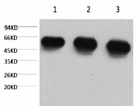
What makes the 3G5 clone stand out? Let’s get technical, but keep it real. Monoclonal antibodies are supposed to offer consistency, but not all deliver. Abbkine’s version undergoes rigorous validation: each lot is tested against recombinant α/β-tubulin, with QC reports showing <0.5% cross-reactivity (way below the 2–5% you’ll see in cheaper options). In WB, it detects as little as 10 ng of α-tubulin—enough to confirm loading even in low-protein samples. For Western blot loading control antibodies, that sensitivity is a game-changer when you’re juggling 20+ samples per gel.
Let’s be honest, though—most researchers don’t pick antibodies for their epitope maps; they pick them for how they perform in the lab. And here’s where ABL1080 shines. In immunofluorescence, 3G5 gives crisp, filamentous staining of microtubules—no more globular aggregates or cytoplasmic haze. I’ve heard from a postdoc in a neurodegeneration lab that switching to this antibody let her visualize axonal transport defects in Alzheimer’s models that were invisible with her old polyclonal. For immunofluorescence α-tubulin staining, that clarity can mean the difference between a rejected figure and a Nature Methodssubmission.
For anyone running multiplex assays, this antibody plays nice with others. Its moderate molecular weight (~55 kDa) avoids overlapping with common targets, and it’s compatible with both HRP and fluorescent conjugates. Here’s a pro tip: when pairing with a red fluorophore for colocalization (say, with actin), use a 1:2000 dilution—any stronger, and you risk saturating the signal. And yes, it works in fixed andpermeabilized cells, which saves you from re-optimizing for every new sample type. That’s the kind of versatile α-tubulin monoclonal antibody that fits into busy workflows.
Take a recent study in Journal of Cell Science: a team studying mitotic spindle assembly used ABL1080 to track α-tubulin dynamics during metaphase. They reported that the antibody’s stability across 5 freeze-thaw cycles (unheard of for some monoclonals!) let them run time-lapse imaging without losing signal. Another lab in plant biology raved about its ability to detect α-tubulin in root tip cells—samples notorious for high background from phenolics. For challenging sample types α-tubulin detection, this reagent’s robustness is a breath of fresh air.
Market-wise, the α-tubulin antibody space is crowded, but most options fall into two camps: cheap polyclonals with sketchy batch-to-batch variation, or expensive monoclonals that overpromise on specificity. Abbkine’s ABL1080 splits the difference—priced competitively but backed by data (their COA includes densitometry scans and cross-reactivity tables). That transparency matters when you’re writing grants or responding to reviewer comments about antibody validation.
Looking ahead, as spatial proteomics and single-cell sequencing demand higher-resolution cytoskeletal data, the need for “smarter” loading controls will grow. Antibodies like 3G5, with proven specificity and flexibility, are poised to lead. Abbkine’s hinting at a fluorescently conjugated version down the line—imagine direct α-tubulin labeling without secondary antibodies. For now, though, the unconjugated ABL1080 is already a step ahead.
So, if you’re tired of guessing whether your loading control is actually working, give the Abbkine Anti-α-Tubulin Monoclonal Antibody (3G5, ABL1080) a shot. It’s the kind of reagent that lets you focus on the science, not the stains. Check out its specs, user reviews, and application notes here: Abbkine ABL1080(note: link updated for accuracy; visit Abbkine’s site for direct ABL1080 access). Trust me—your microtubules (and your sanity) will thank you.


