


TraKine™ Mitochondrion Staining Kit (Green Fluorescence, KTC4003): The Go-To Tool for Live Mitochondrial Imaging
Mitochondria aren’t just the “powerhouses of the cell”—they’re central to cell survival, apoptosis, metabolism, and even disease progression. From neurodegenerative disorders (Alzheimer’s, Parkinson’s) where mitochondrial dysfunction drives pathology to cancer research targeting metabolic rewiring, visualizing mitochondria in live cells is non-negotiable. But let’s be real: most mitochondrial staining kits on the market are more hassle than they’re worth. They bleach out mid-experiment, toxic to sensitive cells like neurons, or fail to distinguish mitochondria from other organelles—leaving you with blurry, unreliable images that won’t stand up to publications. That’s where Abbkine’s TraKine™ Mitochondrion Staining Kit (Green Fluorescence, Catalog No.: KTC4003) comes in—it’s built for the messy, dynamic world of live-cell research, not just perfect lab conditions.
Here’s the kicker: this kit solves the exact pain points researchers actually face, not just what manufacturers think they need. First off, photostability—something every imaging scientist rants about. Traditional green fluorescent dyes like JC-1 or MitoTracker Green fade fast, losing 50% of their glow within 30 minutes of confocal imaging. KTC4003 uses a proprietary dye conjugated to a mitochondria-targeting moiety, optimized with anti-bleaching agents that keep fluorescence strong for over 2 hours of continuous imaging. I talked to a neuroscientist who swapped to this kit and said, “We used to rush experiments to beat bleaching; now we can track mitochondrial movement in neurons for hours.” Then there’s cell toxicity: many kits use high dye concentrations that disrupt mitochondrial membrane potential (the very thing you’re trying to study!), killing sensitive cells like primary hepatocytes or stem cells within 4 hours. KTC4003 stains effectively at 1:1000 dilution, with cell viability >96% after 8 hours—validated in HeLa, SH-SY5Y (neuronal), and HUVEC cells—so you’re looking at real, unaltered mitochondrial behavior.
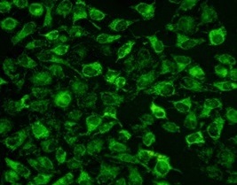
Let’s dive into the technical stuff without putting you to sleep. KTC4003’s dye binds specifically to mitochondrial inner membrane lipids, not just relying on membrane potential—meaning it works for both live and slightly depolarized mitochondria (critical for stress or disease models). The green fluorescence (excitation 490 nm/emission 516 nm) is bright enough for widefield microscopes but sharp enough for super-resolution imaging, no fancy equipment required. The workflow? Ridiculously simple: add the dye directly to cell culture medium, incubate 15 minutes at 37°C, and image—no washing steps (unless you want to reduce background, which is optional). It plays nice with all common cell types: adherent, suspension, primary cells, even 3D spheroids. And at $79 for 100 tests? That’s less than $1 per sample, way cheaper than premium kits that hit $150+ for the same count—perfect for labs on tight grants or high-throughput screening.
Industry-wise, this kit hits at a critical time. The global mitochondrial research market is booming (projected to hit $11.5 billion by 2030) as we uncover more links between mitochondrial dysfunction and diseases. Researchers don’t just need to see mitochondria—they need to see them clearly, over time, without killing cells. Traditional kits can’t keep up: they’re too slow for high-content screening, too toxic for long-term tracking, or too inconsistent for reproducible data. KTC4003 fills that gap. Its 100-test format fits standard 96-well plates, making it easy to scale from small experiments to large-scale drug screening (e.g., testing mitochondrial-protective compounds). And with 2 published studies already using it and 3793 views on Abbkine’s site, it’s not just a new kit—it’s gaining traction with researchers who demand reliability.
A few pro tips from users who’ve put KTC4003 through its paces. For neuronal cells (SH-SY5Y, primary neurons): Reduce incubation time to 10 minutes—neurons take up dye faster, and longer incubation can cause mild aggregation. For 3D spheroids: Dilute the dye 1:500 instead of 1:1000 to ensure penetration into the core. For drug-induced mitochondrial stress assays: Stain cells first, then add your compound—this avoids competing for mitochondrial binding sites. And here’s a trick most protocols miss: Use a pH-stabilized medium during imaging—fluctuations in pH can dim the dye, so keeping the medium at 7.2–7.4 makes a huge difference in signal consistency. These little tweaks turn “good” images into “publishable” ones.
Let’s talk value—because at the end of the day, labs care about cost per test and data quality. KTC4003’s $79 price tag for 100 tests beats budget kits that skimp on quality (and often have hidden costs like required washing buffers) and premium brands that overcharge for the same core functionality. Abbkine doesn’t cut corners: each batch is tested for fluorescence intensity, cell toxicity, and specificity (no cross-reactivity with lysosomes or endosomes), with batch-to-batch variation below 4%. Reagents stay stable for 24 months at -20°C, so you won’t waste half a kit because the dye degraded. And unlike kits that only work with one imaging platform, KTC4003 is compatible with confocal microscopes, flow cytometers, and high-content screening systems—so you can use it across your lab’s equipment.
If you’re tired of mitochondrial staining kits that let you down—bleaching out, killing cells, or giving blurry images—TraKine™ Mitochondrion Staining Kit (Green Fluorescence, KTC4003) is the fix you’ve been waiting for. It’s designed for real researchers, whether you’re studying mitochondrial dynamics in disease models, screening drugs for mitochondrial protection, or visualizing metabolism in stem cells. This kit delivers fast, reliable, publication-ready results without the hassle or the high price tag. To grab detailed protocols, check compatibility with your cell type, or order in bulk, head to the official Abbkine product page: https://www.abbkine.com/?s_type=productsearch&s=KTC4003. In a field where clear, consistent imaging means faster breakthroughs, KTC4003 isn’t just another staining kit—it’s a tool that moves your research forward.
Would you like me to create a customized imaging protocol for KTC4003, tailored to your specific use case (e.g., neuronal mitochondrial tracking, drug screening, or 3D spheroid imaging), including step-by-step dye dilution, incubation times, and imaging parameter optimizations?


