


TraKine™ Cell Plasma Membrane Staining Kit (Orange Fluorescence) (Abbkine KTC4002): A Practical Guide to Specific, Low-Toxic Membrane Visualization
Cell plasma membrane staining is a cornerstone technique in cell biology, enabling visualization of cell morphology, membrane integrity, and interactions in applications ranging from live-cell imaging and flow cytometry to drug delivery studies and cytotoxicity assays. Yet, traditional membrane stains often present critical limitations: green or blue fluorophores suffer from high cellular autofluorescence, toxic reagents induce cell death (hindering long-term tracking), or poor membrane specificity leads to cytoplasmic leakage—compromising data quality. Abbkine’s TraKine™ Cell Plasma Membrane Staining Kit (Orange Fluorescence) (catalog KTC4002, available at https://www.abbkine.com/?s_type=productsearch&s=KTC4002) addresses these pain points with a cell-friendly, specificity-optimized design. Priced at $69 for 50 tests, this kit leverages an orange fluorophore (excitation ~550nm, emission ~570nm) and a membrane-targeting moiety to deliver crisp, non-toxic staining—making it ideal for both live and fixed cell applications. This practical guide offers research-grade strategies to master the kit, from sample-specific staining protocols to imaging optimization, ensuring you unlock its full potential for reliable membrane visualization.
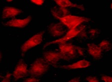
At the core of TraKine™ Cell Plasma Membrane Staining Kit (Orange Fluorescence) KTC4002’s performance lies its proprietary orange fluorophore and membrane-targeting chemistry. Unlike generic lipophilic dyes that non-specifically insert into all lipid bilayers (including organelles), this kit’s fluorophore is conjugated to a cholestanol derivative— a molecule with high affinity for the outer leaflet of the plasma membrane (due to its structural similarity to cholesterol). This targeting ensures minimal internalization into endosomes or mitochondria, eliminating cytoplasmic background that plagues low-cost stains. The orange fluorescence emission is a game-changer for reducing autofluorescence: most cells and tissues exhibit weak autofluorescence in the orange spectrum, compared to the green range (where flavins and NADH emit strongly). For researchers conducting multi-color imaging (e.g., co-staining with GFP-tagged proteins or DAPI for nuclei), this spectral separation prevents signal overlap—critical for quantitative analysis. Additionally, the fluorophore’s high photostability (resisting bleaching for >60 minutes under confocal laser illumination) supports time-lapse imaging of live cells, a key advantage over photolabile dyes that fade within minutes.
Tailoring sample preparation to cell type is foundational to achieving crisp, specific membrane staining with KTC4002. For adherent cells (e.g., HeLa, MCF-7, primary fibroblasts): Seed cells onto coverslips or imaging dishes to 60–70% confluency (overconfluent cells crowd membranes, obscuring details). Wash twice with pre-warmed PBS (pH 7.4) to remove serum components that bind the dye. For suspension cells (e.g., Jurkat, PBMCs): Harvest 1×10⁶ cells by centrifugation (300×g for 5 minutes), resuspend in serum-free medium to avoid non-specific binding, and transfer to a 96-well black plate (for imaging) or flow cytometry tube. For primary cells (e.g., neurons, hepatocytes)—which are more sensitive to dye toxicity: Reduce the final dye concentration by 50% (see optimization below) and use serum-supplemented medium during staining to maintain cell viability. A critical step: Avoid fixing cells before staining if live-cell imaging is intended—formaldehyde crosslinking alters membrane lipid structure, reducing dye binding. For fixed cells: Use 4% paraformaldehyde (PFA) for 10 minutes at room temperature, then permeabilize lightly (0.1% Triton X-100 for 5 minutes) to enhance dye access (skip permeabilization for strict membrane-only staining).
Optimizing staining conditions unlocks KTC4002’s full specificity and minimizes toxicity, especially for long-term or sensitive applications. Start with dye concentration: The kit’s recommended working concentration is 1 μM, but adjust based on cell type: 0.5 μM for primary/sensitive cells, 1–2 μM for immortalized cell lines (e.g., HEK293, CHO). Incubation time and temperature: For live cells, incubate at 37°C for 15–20 minutes (allows uniform membrane insertion); for fixed cells, incubate at room temperature for 10 minutes (avoids unnecessary cell stress). Avoid over-incubation (>30 minutes), as this increases the risk of cytoplasmic leakage—especially in cells with compromised membranes. Washing is critical: After staining, wash cells 3× with PBS (live cells) or staining buffer (fixed cells) to remove unbound dye—residual free dye causes background fluorescence. For multi-color staining: Stain the plasma membrane first with KTC4002, then add nuclear dyes (e.g., DAPI, Hoechst) or target-specific probes (e.g., Alexa Fluor 488-conjugated antibodies) — the orange spectrum (570nm emission) is spectrally distinct from green (520nm) and blue (460nm) channels, preventing crosstalk.
Mastering imaging parameters is essential to capturing high-quality, quantitative data with TraKine™ Cell Plasma Membrane Orange Fluorescence Staining Kit KTC4002. For confocal microscopy: Use a 543nm HeNe laser or 561nm diode laser for excitation, and a 575–620nm emission filter to isolate orange fluorescence. Adjust laser power to 10–20% (to avoid photobleaching) and gain to minimize background—overexposure washes out membrane details. For flow cytometry: Set the excitation to 561nm (PE channel) and emission to 585nm—gate on single cells to exclude debris, and use unstained cells as a negative control to set the threshold for membrane-positive populations. For widefield microscopy: Use a rhodamine filter set (excitation 530–560nm, emission 570–640nm) and ensure uniform illumination to avoid uneven staining artifacts. A pro tip for live-cell time-lapse: Use a temperature-controlled chamber (37°C, 5% CO₂) and image every 5–10 minutes—KTC4002’s low toxicity (cell viability >90% after 24 hours of staining, per Abbkine’s validation) enables long-term tracking of membrane dynamics (e.g., cell migration, membrane blebbing).
Troubleshooting common technical hurdles ensures consistent, publishable results with KTC4002. If cytoplasmic background is high: Reduce dye concentration (e.g., from 2 μM to 1 μM) or shorten incubation time (to 10 minutes); ensure thorough washing (add a fourth PBS wash). If membrane staining is patchy: Verify cell confluency (avoid overcrowding) and ensure the dye is freshly diluted—aliquot the stock solution (1 mM) into small volumes to avoid repeated freeze-thaw cycles (which degrade the fluorophore). If photobleaching occurs quickly: Increase the dye concentration slightly (e.g., 1.5 μM) or use an anti-fade mounting medium (for fixed cells) or imaging buffer with antioxidants (for live cells). If cells die during staining (primary cells): Switch to serum-supplemented medium during incubation, reduce dye concentration, and avoid 37°C incubation (use room temperature for 20 minutes). For multi-color crosstalk: Adjust emission filters to narrow the bandpass (e.g., 580–610nm for KTC4002) or reduce the concentration of the overlapping dye (e.g., GFP-tagged protein).
The versatility of TraKine™ Cell Plasma Membrane Staining Kit (Orange Fluorescence) KTC4002 aligns with the growing demand for multi-purpose membrane visualization tools across research fields. In cell biology, it enables tracking of membrane integrity during drug treatment (e.g., assessing cytotoxicity of chemotherapeutics via membrane blebbing). In immunology, it labels immune cells (e.g., T cells, macrophages) for flow cytometric analysis of cell-cell interactions. In neuroscience, it visualizes neuronal membrane morphology (e.g., dendrites, axons) without disrupting viability—critical for studying synaptic plasticity. In drug delivery, it monitors the uptake of liposomal or nanoparticle carriers by visualizing membrane association. What sets KTC4002 apart is its balance of specificity, low toxicity, and spectral compatibility: unlike green membrane stains (e.g., DiO), it avoids autofluorescence; unlike red stains (e.g., DiI), it is compatible with common GFP-based tools. At a cost-per-test of ~$1.38, it democratizes high-quality membrane staining for academic labs, biotech firms, and drug discovery teams.
Best practices for kit storage and handling extend its lifespan and maintain performance. Store the 1 mM stock dye at -20°C, protected from light (wrap in aluminum foil)—the fluorophore is photosensitive and degrades with exposure to ambient light. Aliquot the stock into 5–10 μL volumes to avoid repeated freeze-thaw cycles, which break down the cholestanol-membrane targeting moiety. The staining buffer can be stored at 4°C for up to 1 month or -20°C for long-term storage. Once diluted to working concentration (0.5–2 μM), use the dye within 2 hours to ensure maximal membrane binding. For long-term projects, track batch numbers and include a positive control (e.g., HEK293 cells stained with the recommended concentration) in every experiment—this validates dye activity and identifies batch-to-batch variability early.
In conclusion, Abbkine’s TraKine™ Cell Plasma Membrane Staining Kit (Orange Fluorescence) KTC4002 delivers the specificity, low toxicity, and spectral flexibility required for reliable membrane visualization in live and fixed cells. By following the practical strategies outlined—tailored sample preparation, optimized staining conditions, imaging parameter fine-tuning, and troubleshooting—you can consistently generate high-quality data that advances your research, whether in cell biology, immunology, neuroscience, or drug discovery. This kit’s user-centric design and academic-grade performance make it an indispensable tool for anyone working with cell membrane visualization. To integrate KTC4002 into your workflow, visit its product page at https://www.abbkine.com/?s_type=productsearch&s=KTC4002 and elevate your membrane staining to publication-quality standards.


