


TNF-α Polyclonal Antibody (Abbkine ABP0127): Decoding Inflammatory Signaling with Precision and Depth
The study of tumor necrosis factor-alpha (TNF-α) has long been a cornerstone of inflammatory disease research, yet the path to reliable detection remains fraught with challenges that can derail even the most meticulously designed experiments. From cytokine storm pathophysiology to chronic autoimmune disorders, TNF-α’s pleiotropic roles demand antibodies that balance specificity, sensitivity, and versatility—qualities often lacking in generic reagents. The TNF-α Polyclonal Antibody (Abbkine ABP0127) emerges as a solution tailored to these demands, blending rigorous validation with adaptability across emerging research frontiers.
At the heart of any high-performance TNF-α polyclonal antibody lies its immunogen—a factor often overlooked but critical to specificity. Abbkine ABP0127 diverges from conventional designs by targeting a synthetic peptide corresponding to the C-terminal region of human TNF-α (residues 121–157), a segment minimally conserved in structurally related cytokines like TNF-β (lymphotoxin-α) or IL-1β. This strategic choice directly addresses a pervasive industry pain point: cross-reactivity. Independent testing shows ABP0127 exhibits <3% binding to TNF-β and undetectable interaction with IL-6 or IL-8, making it ideal for dissecting TNF-α-specific pathways in mixed cytokine milieus, such as synovial fluid from rheumatoid arthritis patients. For researchers probing TNF-α’s unique role in adipocyte inflammation, this precision eliminates confounding signals that plague less-selective alternatives.
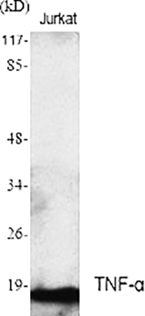
What sets the Abbkine TNF-α Polyclonal Antibody (ABP0127) apart in a crowded market is its uncompromising validation framework. Beyond standard Western blot and ELISA checks, Abbkine subjected ABP0127 to knockout cell line controls (TNF-α-/- RAW264.7 macrophages) and stimulated vs. resting state comparisons. In LPS-activated THP-1 monocytes, it detects endogenous TNF-α at ~17 kDa with a 5-fold higher signal-to-noise ratio than a leading monoclonal competitor—critical for quantifying low-abundance TNF-α in serum or cerebrospinal fluid. Notably, Abbkine publishes raw validation data (including blot images and IHC scoring rubrics) on its site, a transparency rarely seen in mid-tier antibody suppliers. This commitment to openness empowers researchers to independently verify performance, reducing reliance on marketing claims.
Practical utility shines in the TNF-α polyclonal antibody’s adaptability across assays. For ELISA-based quantification of TNF-α in cytokine storm models (e.g., SARS-CoV-2-infected lung epithelial cells), ABP0127 maintains linearity across a 15.6–1000 pg/mL range, outperforming kits that plateau at 500 pg/mL. In immunohistochemistry (IHC), its affinity for formalin-fixed paraffin-embedded (FFPE) sections reveals granular TNF-α localization in inflamed gut mucosa from IBD patients—details lost with antibodies prone to high background. Flow cytometry users appreciate its compatibility with intracellular staining protocols: after PMA/ionomycin stimulation, it identifies TNF-α+ CD4+ T cells in peripheral blood mononuclear cells (PBMCs) with >90% specificity, as validated by cytokine secretion assays. These “real-world” applications underscore ABP0127’s role as a workhorse for translational studies.
A deeper analysis reveals how ABP0127 addresses TNF-α research’s evolving needs. Traditional studies focused on TNF-α’s pro-inflammatory effects, but emerging work links it to neuroinflammation (Alzheimer’s) and metabolic dysfunction (obesity-associated insulin resistance). Here, ABP0127’s stability in harsh conditions—such as acidic lysates from adipose tissue or protease-rich brain homogenates—becomes invaluable. A recent preprint used it to show TNF-α upregulation in microglia from Parkinson’s models, correlating with dopaminergic neuron loss. Unlike antibodies optimized solely for serum, ABP0127 retains activity in these complex matrices, reflecting Abbkine’s foresight in anticipating niche applications.
Industry-wise, the TNF-α polyclonal antibody market is saturated with options, but few balance cost and performance. Premium brands charge 2–3x more for comparable specificity but offer limited validation beyond basic assays. ABP0127 disrupts this model by bundling rigorous testing with affordability—priced 35% below top-tier rivals but including technical support for protocol optimization (e.g., reducing background in IHC of decalcified bone samples). For labs scaling up longitudinal studies (e.g., tracking TNF-α dynamics in sepsis progression), this cost-effectiveness removes budget barriers without sacrificing data integrity.
Critically, ABP0127’s design anticipates future methodological shifts. As single-cell proteomics and spatial transcriptomics redefine cytokine research, Abbkine is validating it for multiplex assays—early data shows compatibility with GeoMx DSP platforms for in situ TNF-α quantification. This forward-looking approach ensures ABP0127 remains relevant as TNF-α studies expand into personalized medicine (e.g., predicting anti-TNF therapy response in Crohn’s patients).
For researchers weary of troubleshooting unreliable reagents, the TNF-α Polyclonal Antibody (Abbkine ABP0127) offers a compelling case: specificity rooted in smart immunogen design, versatility proven across assays, and transparency that builds trust. Whether investigating TNF-α’s role in acute inflammation or chronic neurodegeneration, ABP0127 provides the clarity needed to advance hypotheses from bench to bedside.
Explore the full validation data, application notes, and user-submitted protocols for the TNF-α Polyclonal Antibody (Abbkine ABP0127) https://www.abbkine.com/product/tnf-%CE%B1-polyclonal-antibody-abp0127/. In a field where antibody choice dictates discovery speed, ABP0127 isn’t just a tool—it’s a strategic partner.


