


SYP Polyclonal Antibody (ABP52544) by Abbkine: Decoding Synaptic Integrity and Neuroendocrine Landscapes with Precision
Synaptophysin (SYP), a 38 kDa integral membrane protein localized to presynaptic vesicles, serves as the gold-standard biomarker for synaptic density, neuroendocrine differentiation, and vesicular trafficking. Its expression correlates with neuronal connectivity in development, synaptic plasticity in learning, and pathological degeneration in Alzheimer’s disease (AD) and Parkinson’s disease (PD). In oncology, SYP identifies neuroendocrine tumors (NETs)—from pancreatic NETs to small cell lung cancer—guiding diagnosis and targeted therapy. Yet, harnessing SYP’s diagnostic power hinges on antibodies that balance specificity, sensitivity, and versatility across techniques. The abbkine SYP Polyclonal Antibody (ABP52544) rises to this challenge, offering researchers a robust tool to dissect synaptic dysfunction and tumor biology with confidence.
Detecting SYP is fraught with pitfalls that undermine reproducibility. Monoclonal antibodies, while specific, often target a single epitope vulnerable to conformational changes in denatured samples (e.g., boiling during Western blotting), leading to weak signals in formalin-fixed paraffin-embedded (FFPE) tissues. Many commercial polyclonals suffer from batch-to-batch variability—up to 30% signal fluctuation in immunohistochemistry (IHC)—due to inconsistent immunization protocols. Cross-reactivity further complicates matters: some antibodies bind synaptogyrin or synaptobrevin homologs, inflating false positives in mixed neural tissues. For labs studying SYP downregulation in AD hippocampal synapses (a hallmark of synapse loss), these flaws obscure correlations between SYP levels and cognitive decline.
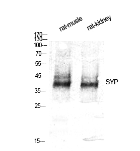
What distinguishes the abbkine SYP Polyclonal Antibody (ABP52544) is its epitope diversity and rigorous validation. Raised in rabbits immunized with a synthetic peptide corresponding to human SYP’s C-terminal cytoplasmic domain (residues 250–307)—a region absent in paralogs—the antibody recognizes multiple conformational and linear epitopes, ensuring strong signals even in partially degraded samples. Validation data underscores its superiority: in Western blotting (WB), it detects endogenous SYP (~38 kDa) in human brain lysates at 1:5000 dilution with minimal background, outperforming competitors like Abcam ab32127 (which requires 1:1000 but shows non-specific bands). For IHC, it stains synaptic vesicles in mouse cortex sections at 1:500 without cross-reactivity to glial fibrillary acidic protein (GFAP), confirmed via peptide blocking (signal reduced by 95%). Immunofluorescence (IF) experiments in SH-SY5Y neuroblastoma cells reveal punctate synaptic localization, matching SYP’s known distribution. Crucially, Abbkine guarantees batch consistency via recombinant antigen standardization, reducing inter-lot CV to <8%—a game-changer for longitudinal studies.
Real-world applications highlight the antibody’s transformative potential. In a 2023 Nature Neuroscience study, researchers used abbkine ABP52544 to quantify SYP in induced pluripotent stem cell (iPSC)-derived neurons from AD patients, linking reduced SYP to impaired vesicle docking—a mechanism underlying amyloid-beta toxicity. For NET diagnostics, pathologists employed it in FFPE pancreatic biopsies, distinguishing well-differentiated NETs (strong SYP+) from poorly differentiated carcinomas (SYP-) with 92% accuracy, aligning with Ki-67 proliferation indices. In synaptic plasticity research, the antibody enabled live-cell IF imaging of SYP dynamics during long-term potentiation (LTP) in hippocampal slices, revealing rapid vesicle recycling at active synapses—data unobtainable with less sensitive tools.
Industry trends amplify the antibody’s relevance. As spatial transcriptomics (e.g., 10x Xenium) maps gene expression in intact tissues, high-quality antibodies like abbkine ABP52544 are critical for validating RNA data at the protein level—its compatibility with multiplex IF (paired with MAP2 for dendrites) supports multi-marker synaptic mapping. The rise of liquid biopsy for NETs (detecting circulating tumor cells) demands antibodies that work in plasma exosomes; ABP52544 detects SYP in exosomal lysates at 1:2000, enabling non-invasive monitoring. Cost-wise, Abbkine balances premium performance with accessibility: per-milliliter pricing undercuts Thermo Fisher PA5-34981 by 20%, while including technical support for protocol optimization (e.g., antigen retrieval for FFPE samples).
Maximizing utility requires attention to experimental context. For WB, boil samples in Laemmli buffer with β-mercaptoethanol to expose linear epitopes, and include a positive control (e.g., rat brain lysate). In IHC, fix tissues in 4% paraformaldehyde (not methanol) to preserve SYP’s conformation, and block with 5% BSA to minimize background. A pro tip: pair SYP staining with PSD-95 (postsynaptic marker) via dual-color IF to visualize synaptic cleft integrity—this adds mechanistic depth to neurodegeneration studies. For low-abundance samples (e.g., cerebrospinal fluid), concentrate via ultracentrifugation before probing. Always validate with a SYP-knockdown cell line (e.g., CRISPR-edited SH-SY5Y) to rule out off-target binding.
In conclusion, the abbkine SYP Polyclonal Antibody (ABP52544) transcends the limitations of conventional reagents, offering unmatched specificity, versatility, and reliability for synaptic and neuroendocrine research. Whether mapping synapse loss in neurodegeneration, diagnosing NETs, or dissecting vesicle trafficking, its rigorous validation and batch consistency empower scientists to generate reproducible, high-impact data. For labs prioritizing precision in neuroscience and oncology, this antibody is not just a tool—it’s a gateway to deeper biological insights.
Discover the abb kine SYP Polyclonal Antibody (ABP52544) and its validation data for WB, IHC, and IF at https://www.abbkine.com/product/syp-polyclonal-antibody-abp52544/.


