| Product name | XRCC4 Monoclonal Antibody |
| Immunogen | Synthetic Peptide |
| Host | Mouse |
| Reactivity | Human |
| Applications | WB,IHC,IF,IP |
| Applications notes | Optimal working dilutions should be determined experimentally by the investigator. Suggested starting dilutions are as follows: WB 1:2000;IP 1:200;IF 1:200;IHC 1:50-300 |
| Clonality | Monoclonal |
| Preparation method | The antibody was affinity-purified from mouse ascites by affinity-chromatography using specific immunogen. |
| Alternative | XRCC4; DNA repair protein XRCC4; X-ray repair cross-complementing protein 4 |
| Formulation | Liquid solution |
| Concentration | 1 mg/ml |
| Molecular weight | 38kD |
| Storage buffer | PBS, pH 7.4, containing 0.5%BSA, 0.02% sodium azide as Preservative and 50% Glycerol. |
| Storage instructions | Stable for one year at -20°C from date of shipment. For maximum recovery of product, centrifuge the original vial after thawing and prior to removing the cap. Aliquot to avoid repeated freezing and thawing. |
| Shipping | Gel pack with blue ice. |
| Precautions | The product listed herein is for research use only and is not intended for use in human or clinical diagnosis. Suggested applications of our products are not recommendations to use our products in violation of any patent or as a license. We cannot be responsible for patent infringements or other violations that may occur with the use of this product. |
| Background | X-ray repair cross complementing 4 encoded by XRCC4 functions together with DNA ligase IV and the DNA-dependent protein kinase in the repair of DNA double-strand breaks. X-ray repair cross complementing 4 plays a role in both non-homologous end joining and the completion of V (D)J recombination. Mutations in XRCC4 can cause short stature, microcephaly, and endocrine dysfunction (SSMED). Alternative splicing generates several transcript variants. |
| Gene ID | 7518 |
| Alternative | XRCC4; DNA repair protein XRCC4; X-ray repair cross-complementing protein 4 |
| Others | The antibody detects endogenous XRCC4 proteins. |
| Accession | Q13426 |
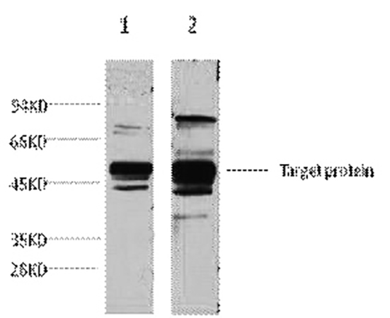
Fig.1. Western blot analysis of 1) Hela, 2) 293T, diluted at 1:3000.
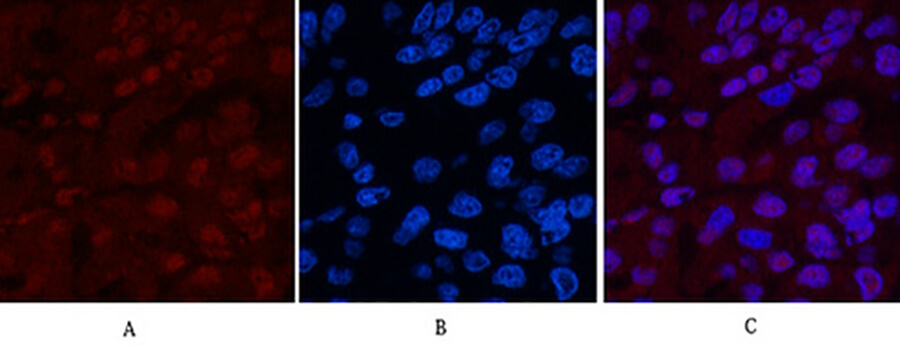
Fig.2. Immunofluorescence analysis of human liver cancer tissue. 1, XRCC4 Monoclonal Antibody (red) was diluted at 1:200 (4°C, overnight). 2, Cy3 Labeled secondary antibody was diluted at 1:300 (room temperature, 50min). 3, Picture B: DAPI (blue) 10min. Picture A: Target. Picture B: DAPI. Picture C: merge of A+B.
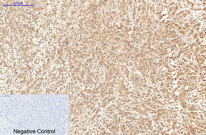
Fig.3. Immunohistochemical analysis of paraffin-embedded human breast cancer tissue. 1, XRCC4 Monoclonal Antibody was diluted at 1:200 (4°C, overnight). 2, Sodium citrate pH 6.0 was used for antibody retrieval (>98°C, 20min). 3, secondary antibody was diluted at 1:200 (room temperature, 30min). Negative control was used by secondary antibody only.
You must be logged in to post a review.


Reviews
There are no reviews yet.