| Product name | TIMP-1 Polyclonal Antibody |
| Immunogen | Synthesized peptide derived from the Internal region of human TIMP-1 at AA range: 30-110 |
| Host | Rabbit |
| Reactivity | Human,Mouse,Rat,Rabbit |
| Applications | IF,WB,IHC,ELISA |
| Applications notes | Optimal working dilutions should be determined experimentally by the investigator. Suggested starting dilutions are as follows: IF 1:50-200;WB 1:500-1:2000;ELISA 1:10000;Not yet tested in other applications; |
| Clonality | Polyclonal |
| Preparation method | The antibody was affinity-purified from rabbit antiserum by affinity-chromatography using epitope-specific immunogen. |
| Alternative | TIMP1; CLGI; TIMP; Metalloproteinase inhibitor 1; Erythroid-potentiating activity; EPA; Fibroblast collagenase inhibitor; Collagenase inhibitor; Tissue inhibitor of metalloproteinases 1; TIMP-1 |
| Formulation | Liquid solution |
| Concentration | 1 mg/ml |
| Molecular weight | 24kD |
| Storage buffer | Liquid in PBS containing 50% glycerol, 0.5% BSA and 0.02% sodium azide. |
| Storage instructions | Stable for one year at -20°C from date of shipment. For maximum recovery of product, centrifuge the original vial after thawing and prior to removing the cap. Aliquot to avoid repeated freezing and thawing. |
| Shipping | Gel pack with blue ice. |
| Precautions | The product listed herein is for research use only and is not intended for use in human or clinical diagnosis. Suggested applications of our products are not recommendations to use our products in violation of any patent or as a license. We cannot be responsible for patent infringements or other violations that may occur with the use of this product. |
| Background | TIMP1 belongs to the TIMP gene family.Metalloproteinase inhibitor 1 encoded by this gene family are natural inhibitors of the matrix metalloproteinases (MMPs), a group of peptidases involved in degradation of the extracellular matrix. In addition to its inhibitory role against most of the known MMPs, the encoded protein is able to promote cell proliferation in a wide range of cell types, and may also have an anti-apoptotic function. Transcription of this gene is highly inducible in response to many cytokines and hormones. In addition, the expression from some but not all inactive X chromosomes suggests that this gene inactivation is polymorphic in human females. TIMP1 is located within intron 6 of the synapsin I gene and is transcribed in the opposite direction. |
| Gene ID | 7076 |
| Alternative | TIMP1; CLGI; TIMP; Metalloproteinase inhibitor 1; Erythroid-potentiating activity; EPA; Fibroblast collagenase inhibitor; Collagenase inhibitor; Tissue inhibitor of metalloproteinases 1; TIMP-1 |
| Others | TIMP-1 Polyclonal Antibody detects endogenous levels of TIMP-1 protein. |
| Accession | P01033 |
| Observed Band(KD) | 24 |
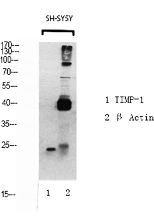
Fig.1. Western Blot analysis of TIMP-1 (1), β-Actin (2), diluted at 1:500.
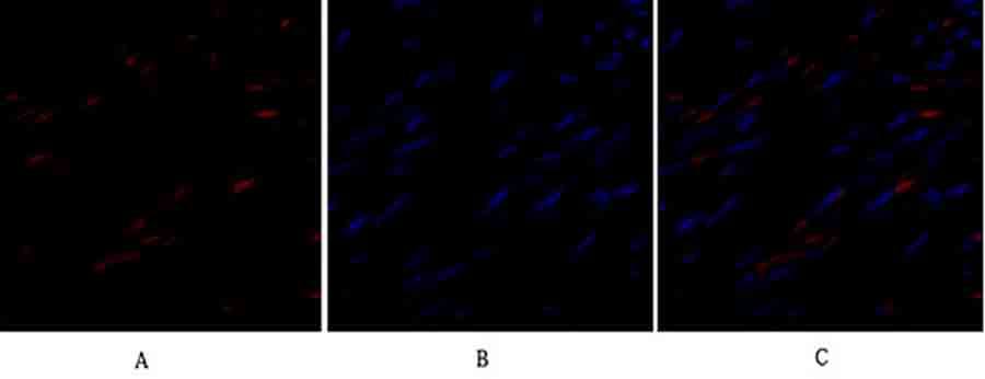
Fig.2. Immunofluorescence analysis of human uterus tissue. 1, TIMP-1 Polyclonal Antibody (red) was diluted at 1:200 (4°C, overnight). 2, Cy3 Labeled secondary antibody was diluted at 1:300 (room temperature, 50min). 3, Picture B: DAPI (blue) 10min. Picture A: Target. Picture B: DAPI. Picture C: merge of A+B.
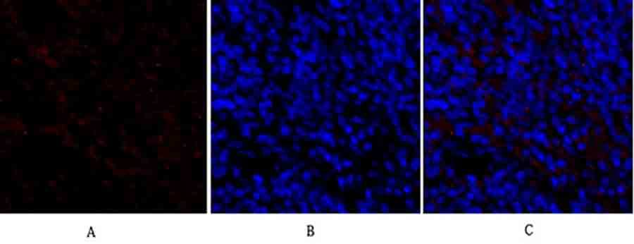
Fig.3. Immunofluorescence analysis of rat spleen tissue. 1, TIMP-1 Polyclonal Antibody (red) was diluted at 1:200 (4°C, overnight). 2, Cy3 Labeled secondary antibody was diluted at 1:300 (room temperature, 50min). 3, Picture B: DAPI (blue) 10min. Picture A: Target. Picture B: DAPI. Picture C: merge of A+B.
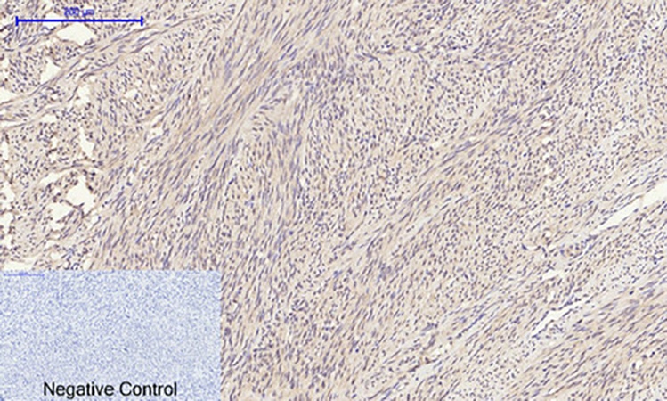
Fig.4. Immunohistochemical analysis of paraffin-embedded human uterus tissue. 1, TIMP-1 Polyclonal Antibody was diluted at 1:200 (4°C, overnight). 2, Sodium citrate pH 6.0 was used for antibody retrieval (>98°C, 20min). 3, secondary antibody was diluted at 1:200 (room temperature, 30min). Negative control was used by secondary antibody only.
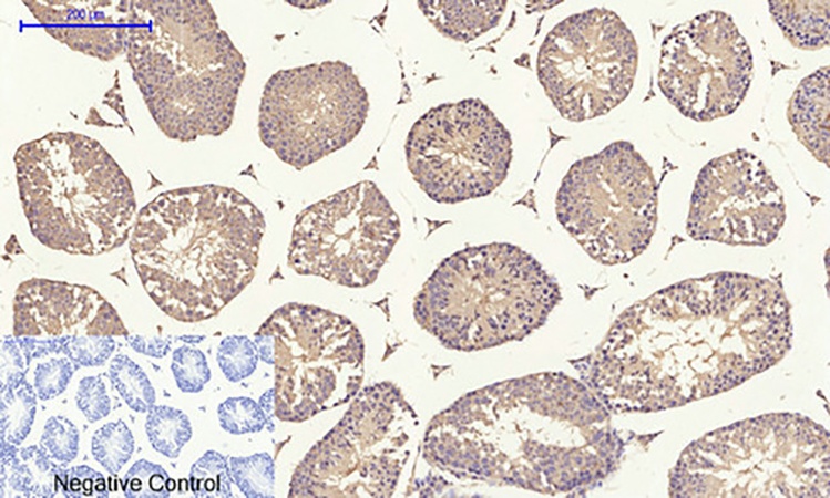
Fig.5. Immunohistochemical analysis of paraffin-embedded mouse testis tissue. 1, TIMP-1 Polyclonal Antibody was diluted at 1:200 (4°C, overnight). 2, Sodium citrate pH 6.0 was used for antibody retrieval (>98°C, 20min). 3, secondary antibody was diluted at 1:200 (room temperature, 30min). Negative control was used by secondary antibody only.
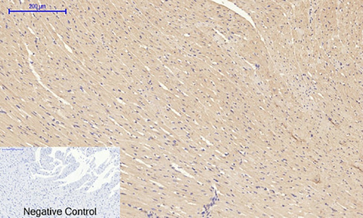
Fig.6. Immunohistochemical analysis of paraffin-embedded rat heart tissue. 1, TIMP-1 Polyclonal Antibody was diluted at 1:200 (4°C, overnight). 2, Sodium citrate pH 6.0 was used for antibody retrieval (>98°C, 20min). 3, secondary antibody was diluted at 1:200 (room temperature, 30min). Negative control was used by secondary antibody only.
You must be logged in to post a review.


Reviews
There are no reviews yet.