| Product name | Tak1 Polyclonal Antibody |
| Immunogen | Synthesized peptide derived from human Tak1 around the non-phosphorylation site of T187 |
| Host | Rabbit |
| Reactivity | Human,Mouse,Rat |
| Applications | IF,WB,IHC,ELISA |
| Applications notes | Optimal working dilutions should be determined experimentally by the investigator. Suggested starting dilutions are as follows: IF 1:50-200;WB 1:500-1:2000;IHC 1:100-1:300;ELISA 1:40000;Not yet tested in other applications; |
| Clonality | Polyclonal |
| Preparation method | The antibody was affinity-purified from rabbit antiserum by affinity-chromatography using epitope-specific immunogen. |
| Alternative | MAP3K7; TAK1; Mitogen-activated protein kinase kinase kinase 7; Transforming growth factor-beta-activated kinase 1; TGF-beta-activated kinase 1 |
| Formulation | Liquid solution |
| Concentration | 1 mg/ml |
| Molecular weight | 77kD |
| Storage buffer | Liquid in PBS containing 50% glycerol, 0.5% BSA and 0.02% sodium azide. |
| Storage instructions | Stable for one year at -20°C from date of shipment. For maximum recovery of product, centrifuge the original vial after thawing and prior to removing the cap. Aliquot to avoid repeated freezing and thawing. |
| Shipping | Gel pack with blue ice. |
| Precautions | The product listed herein is for research use only and is not intended for use in human or clinical diagnosis. Suggested applications of our products are not recommendations to use our products in violation of any patent or as a license. We cannot be responsible for patent infringements or other violations that may occur with the use of this product. |
| Background | Mitogen-activated protein kinase kinase kinase 7 encoded by MAP3K7 is a member of the serine/threonine protein kinase family. This kinase mediates the signaling transduction induced by TGF beta and morphogenetic protein (BMP), and controls a variety of cell functions including transcription regulation and apoptosis. In response to IL-1, this protein forms a kinase complex including TRAF6, MAP3K7P1/TAB1 and MAP3K7P2/TAB2; this complex is required for the activation of nuclear factor kappa B. This kinase can also activate MAPK8/JNK, MAP2K4/MKK4, and thus plays a role in the cell response to environmental stresses. Four alternatively spliced transcript variants encoding distinct isoforms have been reported. |
| Gene ID | 6885 |
| Alternative | MAP3K7; TAK1; Mitogen-activated protein kinase kinase kinase 7; Transforming growth factor-beta-activated kinase 1; TGF-beta-activated kinase 1 |
| Others | Tak1 Polyclonal Antibody detects endogenous levels of Tak1 protein. |
| Accession | O43318 |
| Observed Band(KD) | 70 |
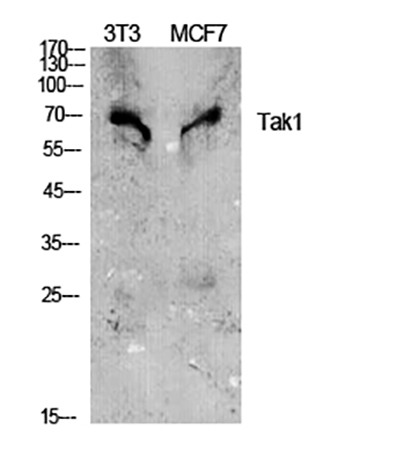
Fig.1. Western Blot analysis of 3T3 (1), MCF-7 (2), diluted at 1:1000.
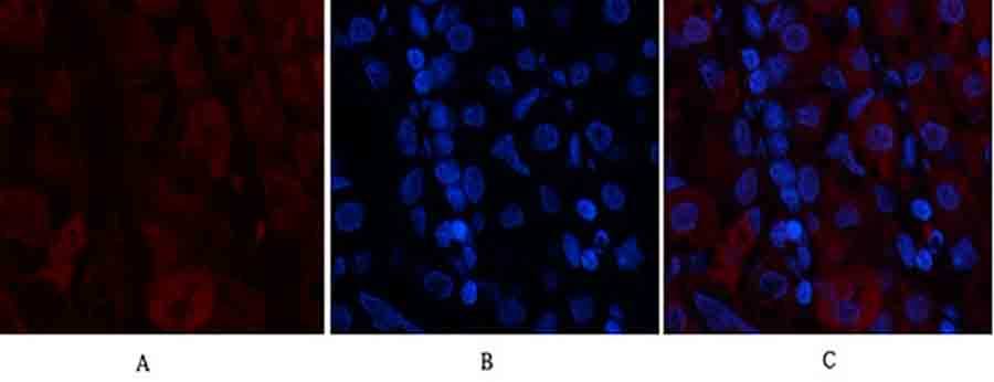
Fig.2. Immunofluorescence analysis of human stomach tissue. 1, Tak1 Polyclonal Antibody (red) was diluted at 1:200 (4°C, overnight). 2, Cy3 Labeled secondary antibody was diluted at 1:300 (room temperature, 50min). 3, Picture B: DAPI (blue) 10min. Picture A: Target. Picture B: DAPI. Picture C: merge of A+B.
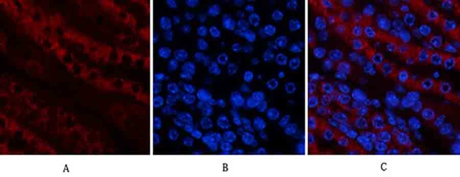
Fig.3. Immunofluorescence analysis of mouse kidney tissue. 1, Tak1 Polyclonal Antibody (red) was diluted at 1:200 (4°C, overnight). 2, Cy3 Labeled secondary antibody was diluted at 1:300 (room temperature, 50min). 3, Picture B: DAPI (blue) 10min. Picture A: Target. Picture B: DAPI. Picture C: merge of A+B.
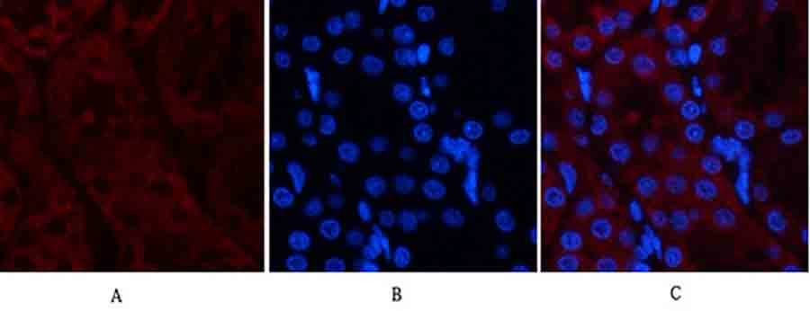
Fig.4. Immunofluorescence analysis of rat kidney tissue. 1, Tak1 Polyclonal Antibody (red) was diluted at 1:200 (4°C, overnight). 2, Cy3 Labeled secondary antibody was diluted at 1:300 (room temperature, 50min). 3, Picture B: DAPI (blue) 10min. Picture A: Target. Picture B: DAPI. Picture C: merge of A+B.
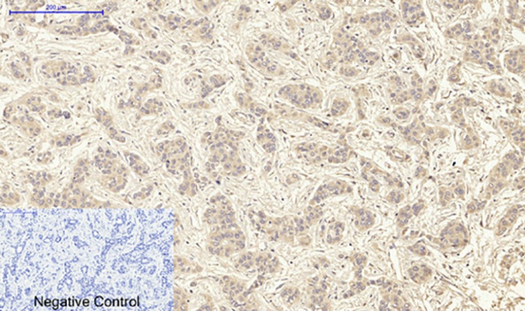
Fig.5. Immunohistochemical analysis of paraffin-embedded human liver cancer tissue. 1, Tak1 Polyclonal Antibody was diluted at 1:200 (4°C, overnight). 2, Sodium citrate pH 6.0 was used for antibody retrieval (>98°C, 20min). 3, secondary antibody was diluted at 1:200 (room temperature, 30min). Negative control was used by secondary antibody only.
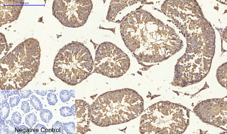
Fig.6. Immunohistochemical analysis of paraffin-embedded mouse testis tissue. 1, Tak1 Polyclonal Antibody was diluted at 1:200 (4°C, overnight). 2, Sodium citrate pH 6.0 was used for antibody retrieval (>98°C, 20min). 3, secondary antibody was diluted at 1:200 (room temperature, 30min). Negative control was used by secondary antibody only.
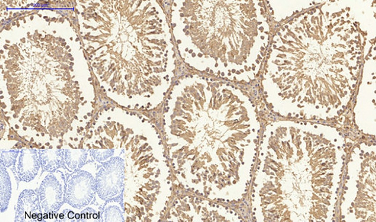
Fig.7. Immunohistochemical analysis of paraffin-embedded rat testis tissue. 1, Tak1 Polyclonal Antibody was diluted at 1:200 (4°C, overnight). 2, Sodium citrate pH 6.0 was used for antibody retrieval (>98°C, 20min). 3, secondary antibody was diluted at 1:200 (room temperature, 30min). Negative control was used by secondary antibody only.
You must be logged in to post a review.


Reviews
There are no reviews yet.