| Product name | SDF-1 Polyclonal Antibody |
| Immunogen | Synthesized peptide derived from the C-terminal region of human SDF-1 |
| Host | Rabbit |
| Reactivity | Human,Mouse,Rat |
| Applications | IF,WB,IHC,ELISA |
| Applications notes | Optimal working dilutions should be determined experimentally by the investigator. Suggested starting dilutions are as follows: IF 1:50-200;WB 1:500-1:2000;IHC 1:100-1:300;ELISA 1:40000;Not yet tested in other applications; |
| Clonality | Polyclonal |
| Preparation method | The antibody was affinity-purified from rabbit antiserum by affinity-chromatography using epitope-specific immunogen. |
| Alternative | CXCL12; SDF1; SDF1A; SDF1B; Stromal cell-derived factor 1; SDF-1; hSDF-1; C-X-C motif chemokine 12; Intercrine reduced in hepatomas; IRH; hIRH; Pre-B cell growth-stimulating factor; PBSF |
| Formulation | Liquid solution |
| Concentration | 1 mg/ml |
| Molecular weight | 10-20kD, |
| Storage buffer | Liquid in PBS containing 50% glycerol, 0.5% BSA and 0.02% sodium azide. |
| Storage instructions | Stable for one year at -20°C from date of shipment. For maximum recovery of product, centrifuge the original vial after thawing and prior to removing the cap. Aliquot to avoid repeated freezing and thawing. |
| Shipping | Gel pack with blue ice. |
| Precautions | The product listed herein is for research use only and is not intended for use in human or clinical diagnosis. Suggested applications of our products are not recommendations to use our products in violation of any patent or as a license. We cannot be responsible for patent infringements or other violations that may occur with the use of this product. |
| Background | CXCL12 encodes a stromal cell-derived alpha chemokine member of the intercrine family. The Stromal cell-derived factor 1 functions as the ligand for the G-protein coupled receptor, chemokine (C-X-C motif) receptor 4, and plays a role in many diverse cellular functions, including embryogenesis, immune surveillance, inflammation response, tissue homeostasis, and tumor growth and metastasis. Mutations in this gene are associated with resistance to human immunodeficiency virus type 1 infections. Multiple transcript variants encoding different isoforms have been found for this gene. |
| Gene ID | 6387 |
| Alternative | CXCL12; SDF1; SDF1A; SDF1B; Stromal cell-derived factor 1; SDF-1; hSDF-1; C-X-C motif chemokine 12; Intercrine reduced in hepatomas; IRH; hIRH; Pre-B cell growth-stimulating factor; PBSF |
| Others | SDF-1 Polyclonal Antibody detects endogenous levels of SDF-1 protein. |
| Accession | P48061 |
| Observed Band(KD) | 10 |
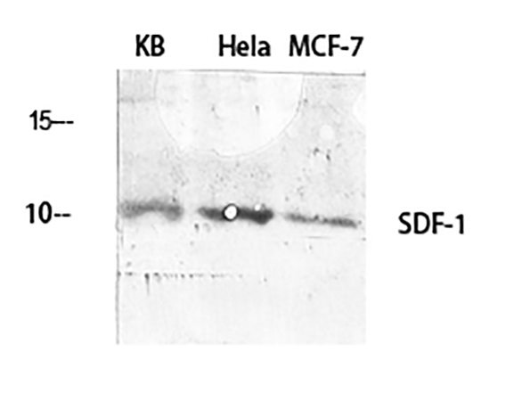
Fig.1. Western Blot analysis of KB (1), Hela (2), MCF-7 (3), diluted at 1:2000.
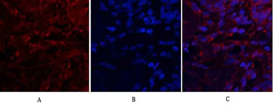
Fig.2. Immunofluorescence analysis of human lung tissue. 1, SDF-1 Polyclonal Antibody (red) was diluted at 1:200 (4°C, overnight). 2, Cy3 Labeled secondary antibody was diluted at 1:300 (room temperature, 50min). 3, Picture B: DAPI (blue) 10min. Picture A: Target. Picture B: DAPI. Picture C: merge of A+B.
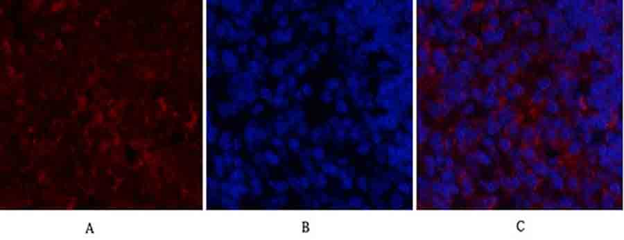
Fig.3. Immunofluorescence analysis of rat spleen tissue. 1, SDF-1 Polyclonal Antibody (red) was diluted at 1:200 (4°C, overnight). 2, Cy3 Labeled secondary antibody was diluted at 1:300 (room temperature, 50min). 3, Picture B: DAPI (blue) 10min. Picture A: Target. Picture B: DAPI. Picture C: merge of A+B.
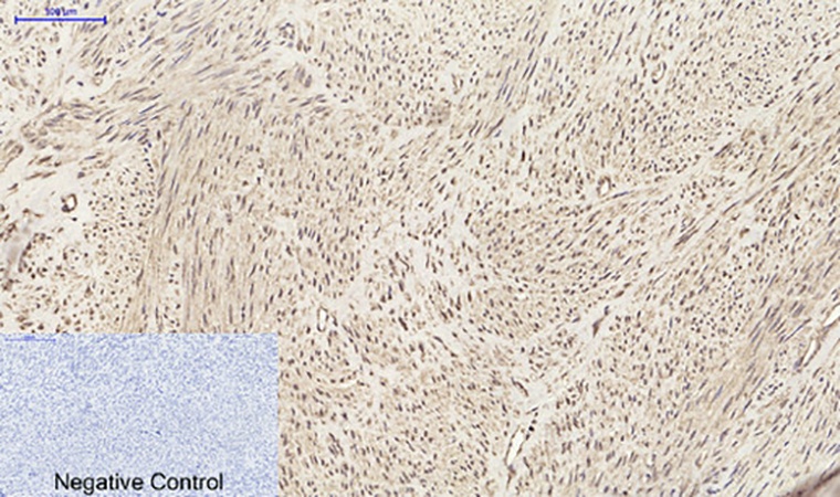
Fig.4. Immunohistochemical analysis of paraffin-embedded human uterus tissue. 1, SDF-1 Polyclonal Antibody was diluted at 1:200 (4°C, overnight). 2, Sodium citrate pH 6.0 was used for antibody retrieval (>98°C, 20min). 3, secondary antibody was diluted at 1:200 (room temperature, 30min). Negative control was used by secondary antibody only.
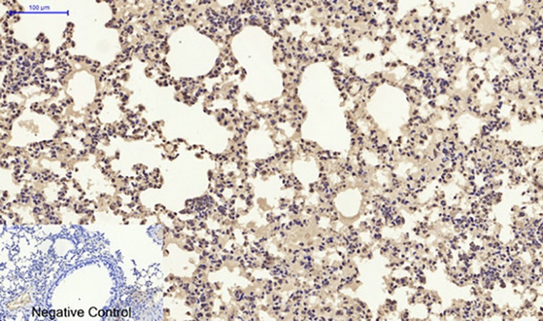
Fig.5. Immunohistochemical analysis of paraffin-embedded mouse lung tissue. 1, SDF-1 Polyclonal Antibody was diluted at 1:200 (4°C, overnight). 2, Sodium citrate pH 6.0 was used for antibody retrieval (>98°C, 20min). 3, secondary antibody was diluted at 1:200 (room temperature, 30min). Negative control was used by secondary antibody only.
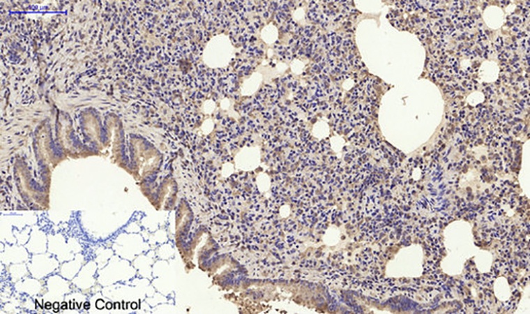
Fig.6. Immunohistochemical analysis of paraffin-embedded rat lung tissue. 1, SDF-1 Polyclonal Antibody was diluted at 1:200 (4°C, overnight). 2, Sodium citrate pH 6.0 was used for antibody retrieval (>98°C, 20min). 3, secondary antibody was diluted at 1:200 (room temperature, 30min). Negative control was used by secondary antibody only.
You must be logged in to post a review.


Reviews
There are no reviews yet.