| Product name | LC3A Mouse Monoclonal Antibody (5G10) |
| Immunogen | Synthetic Peptide of LC3A |
| Host | Mouse |
| Reactivity | Human,Rat,Mouse |
| Applications | WB,IF,IHC |
| Applications notes | Optimal working dilutions should be determined experimentally by the investigator. Suggested starting dilutions are as follows: WB 1:1000-2000;IHC 1:100-200;IF 1:200 |
| Clonality | Monoclonal |
| Preparation method | The antibody was affinity-purified from mouse ascites by affinity-chromatography using specific immunogen. |
| Alternative | Microtubule-associated proteins 1A/1B light chain 3A; Autophagy-related protein LC3 A; Autophagy-related ubiquitin-like modifier LC3 A; MAP1 light chain 3-like protein 1; MAP1A/MAP1B light chain 3 A; MAP1A/MAP1B LC3 A; Microtubule-associated protein 1 light chain 3 alpha |
| Formulation | Liquid solution |
| Concentration | 1 mg/ml |
| Molecular weight | 14,16kD |
| Storage buffer | Liquid in PBS containing 50% glycerol, 0.5% BSA and 0.02% sodium azide. |
| Storage instructions | Stable for one year at -20°C from date of shipment. For maximum recovery of product, centrifuge the original vial after thawing and prior to removing the cap. Aliquot to avoid repeated freezing and thawing. |
| Shipping | Gel pack with blue ice. |
| Precautions | The product listed herein is for research use only and is not intended for use in human or clinical diagnosis. Suggested applications of our products are not recommendations to use our products in violation of any patent or as a license. We cannot be responsible for patent infringements or other violations that may occur with the use of this product. |
| Background | MAP1A and MAP1B are microtubule-associated proteins which mediate the physical interactions between microtubules and components of the cytoskeleton. MAP1A and MAP1B each consist of a heavy chain subunit and multiple light chain subunits. The protein encoded by MAP1LC3A (microtubule associated protein 1 light chain 3 alpha) is one of the light chain subunits and can associate with either MAP1A or MAP1B. Two transcript variants encoding different isoforms have been found for MAP1LC3A. The expression of variant 1 is suppressed in many tumor cell lines, suggesting that may be involved in carcinogenesis. |
| Gene ID | 84557 |
| Alternative | Microtubule-associated proteins 1A/1B light chain 3A; Autophagy-related protein LC3 A; Autophagy-related ubiquitin-like modifier LC3 A; MAP1 light chain 3-like protein 1; MAP1A/MAP1B light chain 3 A; MAP1A/MAP1B LC3 A; Microtubule-associated protein 1 light chain 3 alpha |
| Others | LC3A protein detects endogenous levels of LC3A. |
| Accession | Q9H492 |
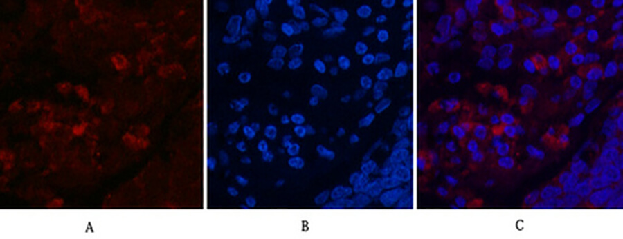
Fig.1. Immunofluorescence analysis of human lung cancer tissue. 1, LC3A Mouse Monoclonal Antibody (5G10) (red) was diluted at 1:200 (4°C, overnight). 2, Cy3 Labeled secondary antibody was diluted at 1:300 (room temperature, 50min). 3, Picture B: DAPI (blue) 10min. Picture A: Target. Picture B: DAPI. Picture C: merge of A+B.
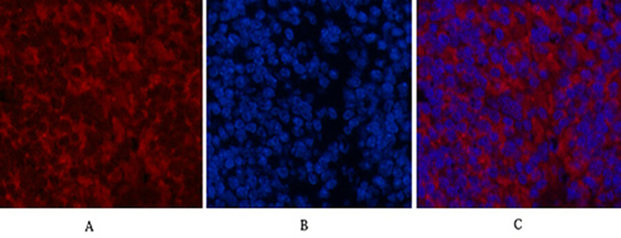
Fig.2. Immunofluorescence analysis of mouse spleen tissue. 1, LC3A Mouse Monoclonal Antibody (5G10) (red) was diluted at 1:200 (4°C, overnight). 2, Cy3 Labeled secondary antibody was diluted at 1:300 (room temperature, 50min). 3, Picture B: DAPI (blue) 10min. Picture A: Target. Picture B: DAPI. Picture C: merge of A+B.
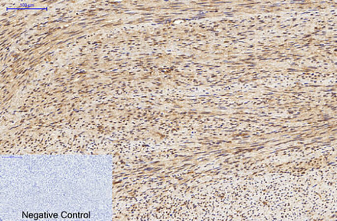
Fig.3. Immunohistochemical analysis of paraffin-embedded human uterus tissue. 1, LC3A Mouse Monoclonal Antibody (5G10) was diluted at 1:200 (4°C, overnight). 2, Sodium citrate pH 6.0 was used for antibody retrieval (>98°C, 20min). 3, secondary antibody was diluted at 1:200 (room temperature, 30min). Negative control was used by secondary antibody only.
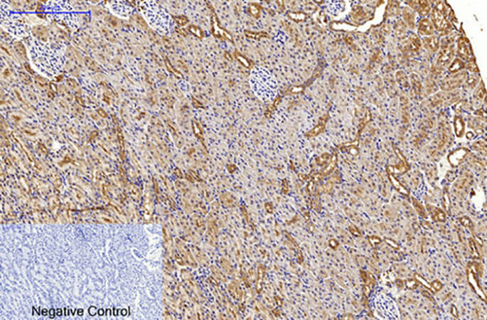
Fig.4. Immunohistochemical analysis of paraffin-embedded rat kidney tissue. 1, LC3A Mouse Monoclonal Antibody (5G10) was diluted at 1:200 (4°C, overnight). 2, Sodium citrate pH 6.0 was used for antibody retrieval (>98°C, 20min). 3, secondary antibody was diluted at 1:200 (room temperature, 30min). Negative control was used by secondary antibody only.
Author:B Chen, B Yang, J Zhu Publication name:International Journal of Molecular sciences IF:4.32
You must be logged in to post a review.


Reviews
There are no reviews yet.