| Product name | HAO1 Monoclonal Antibody |
| Immunogen | Recombinant Protein |
| Host | Mouse |
| Reactivity | Mouse,Rat |
| Applications | WB,IHC,IF, |
| Applications notes | Optimal working dilutions should be determined experimentally by the investigator. Suggested starting dilutions are as follows: WB 1:1000-2000;IF 1:200;IHC 1:50-300 |
| Clonality | Monoclonal |
| Preparation method | The antibody was affinity-purified from mouse ascites by affinity-chromatography using epitope-specific immunogen. |
| Formulation | Liquid solution |
| Concentration | 1 mg/ml |
| Molecular weight | 41kD |
| Storage buffer | PBS, pH 7.4, containing 0.5%BSA, 0.02% sodium azide as Preservative and 50% Glycerol. |
| Storage instructions | Stable for one year at -20°C from date of shipment. For maximum recovery of product, centrifuge the original vial after thawing and prior to removing the cap. Aliquot to avoid repeated freezing and thawing. |
| Shipping | Gel pack with blue ice. |
| Precautions | The product listed herein is for research use only and is not intended for use in human or clinical diagnosis. Suggested applications of our products are not recommendations to use our products in violation of any patent or as a license. We cannot be responsible for patent infringements or other violations that may occur with the use of this product. |
| Background | HAO1 (hydroxyacid oxidase 1) is one of three related genes that have 2-hydroxyacid oxidase activity yet differ in encoded protein amino acid sequence, tissue expression and substrate preference. Subcellular location of the encoded protein is the peroxisome. Specifically, HAO1 is expressed primarily in liver and pancreas and the encoded protein is most active on glycolate, a two-carbon substrate. The protein is also active on 2-hydroxy fatty acids. The transcript detected at high levels in pancreas may represent an alternatively spliced form or the use of a multiple near-consensus upstream polyadenylation site. |
| Gene ID | 54363 |
| Others | The antibody detects endogenous HAO1 protein. |
| Accession | Q9UJM8 |
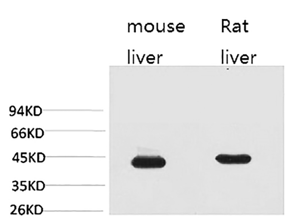
Fig.1. Western blot analysis of Mouse Liver (1), Rat Liver (2).
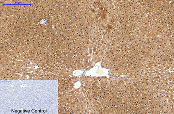
Fig.4. Immunohistochemical analysis of paraffin-embedded rat liver tissue. 1, HAO1 Monoclonal Antibody was diluted at 1:200 (4°C, overnight). 2, Sodium citrate pH 6.0 was used for antibody retrieval (>98°C, 20min). 3, secondary antibody was diluted at 1:200 (room temperature, 30min). Negative control was used by secondary antibody only.
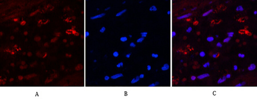
Fig.5. Immunofluorescence analysis of human appendix tissue. 1, HAO1 Monoclonal Antibody (red) was diluted at 1:200 (4°C, overnight). 2, Cy3 Labeled secondary antibody was diluted at 1:300 (room temperature, 50min). 3, Picture B: DAPI (blue) 10min. Picture A: Target. Picture B: DAPI. Picture C: merge of A+B.
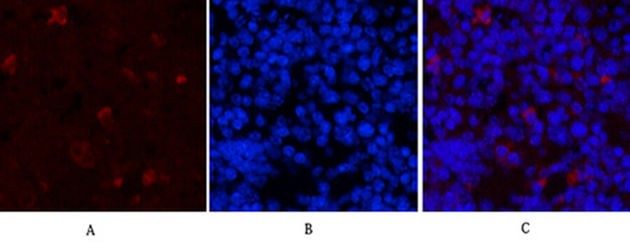
Fig.6. Immunofluorescence analysis of mouse spleen tissue. 1, HAO1 Monoclonal Antibody (red) was diluted at 1:200 (4°C, overnight). 2, Cy3 Labeled secondary antibody was diluted at 1:300 (room temperature, 50min). 3, Picture B: DAPI (blue) 10min. Picture A: Target. Picture B: DAPI. Picture C: merge of A+B.
You must be logged in to post a review.


Reviews
There are no reviews yet.