| Product name | FAS-L Polyclonal Antibody |
| Immunogen | Synthesized peptide derived from the Internal region of human FAS-L at AA range: 70-150 |
| Host | Rabbit |
| Reactivity | Human,Mouse,Pig |
| Applications | WB,IHC,IF,ELISA |
| Applications notes | Optimal working dilutions should be determined experimentally by the investigator. Suggested starting dilutions are as follows: WB 1:500-1:2000;IHC 1:100-1:300;IF 1:200-1:1000;ELISA 1:40000;Not yet tested in other applications. |
| Clonality | Polyclonal |
| Preparation method | The antibody was affinity-purified from rabbit antiserum by affinity-chromatography using epitope-specific immunogen. |
| Alternative | FASLG; APT1LG1; CD95L; FASL; TNFSF6; Tumor necrosis factor ligand superfamily member 6; Apoptosis antigen ligand; APTL; CD95 ligand; CD95-L; Fas antigen ligand; Fas ligand; FasL; CD antigen CD178 |
| Formulation | Liquid solution |
| Concentration | 1 mg/ml |
| Molecular weight | 33kD |
| Storage buffer | Liquid in PBS containing 50% glycerol, 0.5% BSA and 0.02% sodium azide. |
| Storage instructions | Stable for one year at -20°C from date of shipment. For maximum recovery of product, centrifuge the original vial after thawing and prior to removing the cap. Aliquot to avoid repeated freezing and thawing. |
| Shipping | Gel pack with blue ice. |
| Precautions | The product listed herein is for research use only and is not intended for use in human or clinical diagnosis. Suggested applications of our products are not recommendations to use our products in violation of any patent or as a license. We cannot be responsible for patent infringements or other violations that may occur with the use of this product. |
| Background | FASLG is a member of the tumor necrosis factor superfamily. The primary function of tumor necrosis factor ligand superfamily member 6 is the induction of apoptosis triggered by binding to FAS. The FAS/FASLG signaling pathway is essential for immune system regulation, including activation-induced cell death (AICD) of T cells and cytotoxic T lymphocyte induced cell death. It has also been implicated in the progression of several cancers. Defects in this gene may be related to some cases of systemic lupus erythematosus (SLE). Alternatively spliced transcript variants have been described. |
| Gene ID | 356 |
| Alternative | FASLG; APT1LG1; CD95L; FASL; TNFSF6; Tumor necrosis factor ligand superfamily member 6; Apoptosis antigen ligand; APTL; CD95 ligand; CD95-L; Fas antigen ligand; Fas ligand; FasL; CD antigen CD178 |
| Others | FAS-L Polyclonal Antibody detects endogenous levels of FAS-L protein. |
| Accession | P48023 |
| Observed Band(KD) | 33 |
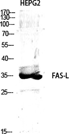
Fig.1. Western Blot analysis of various cells using FAS-L Polyclonal Antibody diluted at 1:1000.
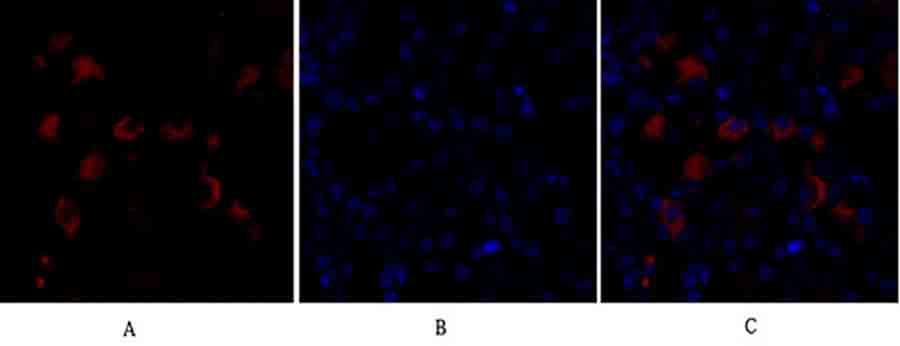
Fig.2. Immunofluorescence analysis of mouse kidney tissue. 1, FAS-L Polyclonal Antibody (red) was diluted at 1:200 (4°C, overnight). 2, Cy3 Labeled secondary antibody was diluted at 1:300 (room temperature, 50min). 3, Picture B: DAPI (blue) 10min. Picture A: Target. Picture B: DAPI. Picture C: merge of A+B.
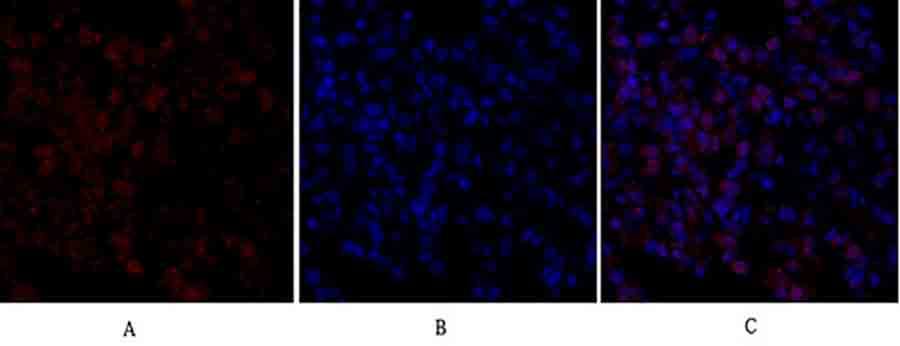
Fig.3. Immunofluorescence analysis of rat lung tissue. 1, FAS-L Polyclonal Antibody (red) was diluted at 1:200 (4°C, overnight). 2, Cy3 Labeled secondary antibody was diluted at 1:300 (room temperature, 50min). 3, Picture B: DAPI (blue) 10min. Picture A: Target. Picture B: DAPI. Picture C: merge of A+B.
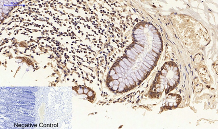
Fig.4. Immunohistochemical analysis of paraffin-embedded human colon tissue. 1, FAS-L Polyclonal Antibody was diluted at 1:200 (4°C, overnight). 2, Sodium citrate pH 6.0 was used for antibody retrieval (>98°C, 20min). 3, secondary antibody was diluted at 1:200 (room temperature, 30min). Negative control was used by secondary antibody only.
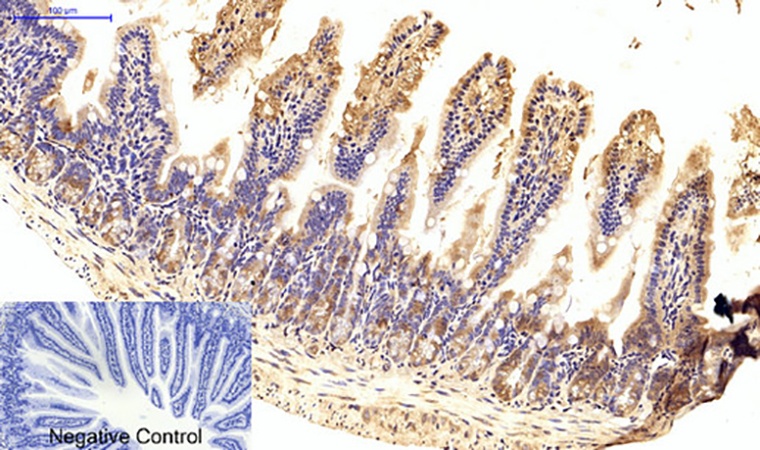
Fig.5. Immunohistochemical analysis of paraffin-embedded mouse colon tissue. 1, FAS-L Polyclonal Antibody was diluted at 1:200 (4°C, overnight). 2, Sodium citrate pH 6.0 was used for antibody retrieval (>98°C, 20min). 3, secondary antibody was diluted at 1:200 (room temperature, 30min). Negative control was used by secondary antibody only.
You must be logged in to post a review.


Reviews
There are no reviews yet.