| Product name | EPAS-1 Polyclonal Antibody |
| Immunogen | Synthesized peptide derived from human EPAS-1 around the non-acetylation site of K385 |
| Host | Rabbit |
| Reactivity | Human,Mouse,Rat |
| Applications | IF,WB,IHC,ELISA |
| Applications notes | Optimal working dilutions should be determined experimentally by the investigator. Suggested starting dilutions are as follows: IF 1:50-200;WB 1:500-1:2000;IHC: 1:100-1:300;ELISA 1:20000;Not yet tested in other applications; |
| Clonality | Polyclonal |
| Preparation method | The antibody was affinity-purified from rabbit antiserum by affinity-chromatography using epitope-specific immunogen. |
| Alternative | EPAS1; BHLHE73; HIF2A; MOP2; PASD2; Endothelial PAS domain-containing protein 1; EPAS-1; Basic-helix-loop-helix-PAS protein MOP2; Class E basic helix-loop-helix protein 73; bHLHe73;HIF-1-alpha-like factor; HLF; Hypoxia-inducible factor 2-alpha; HIF-2-alpha; HIF2-alpha; Member of PAS protein 2; PAS domain-containing protein 2 |
| Formulation | Liquid solution |
| Concentration | 1 mg/ml |
| Molecular weight | 110-120kD |
| Storage buffer | Liquid in PBS containing 50% glycerol, 0.5% BSA and 0.02% sodium azide. |
| Storage instructions | Stable for one year at -20°C from date of shipment. For maximum recovery of product, centrifuge the original vial after thawing and prior to removing the cap. Aliquot to avoid repeated freezing and thawing. |
| Shipping | Gel pack with blue ice. |
| Precautions | The product listed herein is for research use only and is not intended for use in human or clinical diagnosis. Suggested applications of our products are not recommendations to use our products in violation of any patent or as a license. We cannot be responsible for patent infringements or other violations that may occur with the use of this product. |
| Background | EPAS1 encodes a transcription factor involved in the induction of genes regulated by oxygen, which is induced as oxygen levels fall. The endothelial PAS domain protein 1 contains a basic-helix-loop-helix domain protein dimerization domain as well as a domain found in proteins in signal transduction pathways which respond to oxygen levels. Mutations in this gene are associated with erythrocytosis familial type 4. |
| Gene ID | 2034 |
| Alternative | EPAS1; BHLHE73; HIF2A; MOP2; PASD2; Endothelial PAS domain-containing protein 1; EPAS-1; Basic-helix-loop-helix-PAS protein MOP2; Class E basic helix-loop-helix protein 73; bHLHe73;HIF-1-alpha-like factor; HLF; Hypoxia-inducible factor 2-alpha; HIF-2-alpha; HIF2-alpha; Member of PAS protein 2; PAS domain-containing protein 2 |
| Others | EPAS-1 Polyclonal Antibody detects endogenous levels of EPAS-1 protein. |
| Accession | Q99814 |
| Observed Band(KD) | 96 |
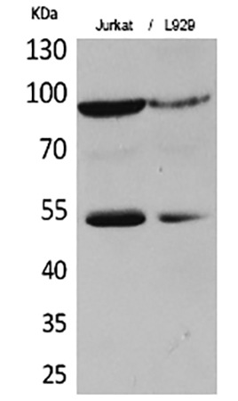
Fig.1. Western Blot analysis of Jurkat (1), L929 (2), diluted at 1:500.
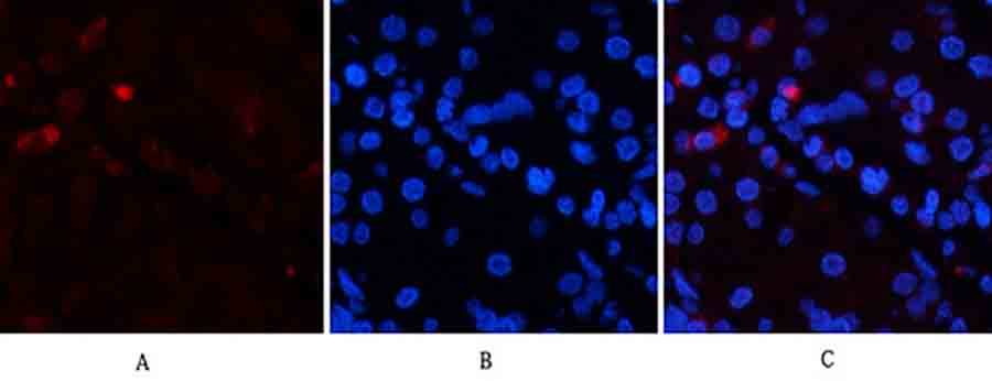
Fig.2. Immunofluorescence analysis of human stomach tissue. 1, EPAS-1 Polyclonal Antibody (red) was diluted at 1:200 (4°C, overnight). 2, Cy3 Labeled secondary antibody was diluted at 1:300 (room temperature, 50min). 3, Picture B: DAPI (blue) 10min. Picture A: Target. Picture B: DAPI. Picture C: merge of A+B.
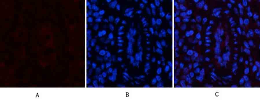
Fig.3. Immunofluorescence analysis of mouse spleen tissue. 1, EPAS-1 Polyclonal Antibody (red) was diluted at 1:200 (4°C, overnight). 2, Cy3 Labeled secondary antibody was diluted at 1:300 (room temperature, 50min). 3, Picture B: DAPI (blue) 10min. Picture A: Target. Picture B: DAPI. Picture C: merge of A+B.
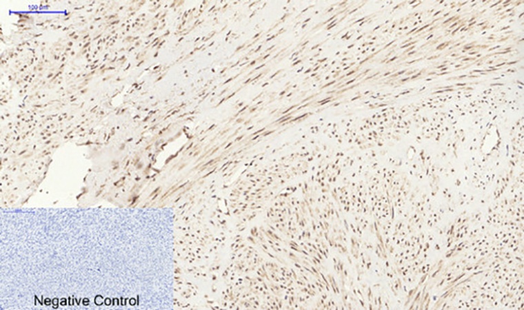
Fig.4. Immunohistochemical analysis of paraffin-embedded human uterus tissue. 1, EPAS-1 Polyclonal Antibody was diluted at 1:200 (4°C, overnight). 2, Sodium citrate pH 6.0 was used for antibody retrieval (>98°C, 20min). 3, secondary antibody was diluted at 1:200 (room temperature, 30min). Negative control was used by secondary antibody only.
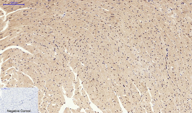
Fig.5. Immunohistochemical analysis of paraffin-embedded mouse heart tissue. 1, EPAS-1 Polyclonal Antibody was diluted at 1:200 (4°C, overnight). 2, Sodium citrate pH 6.0 was used for antibody retrieval (>98°C, 20min). 3, secondary antibody was diluted at 1:200 (room temperature, 30min). Negative control was used by secondary antibody only.
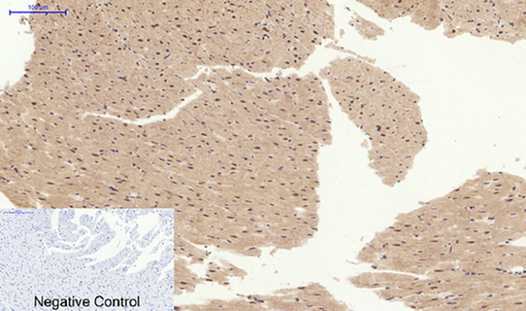
Fig.6. Immunohistochemical analysis of paraffin-embedded rat heart tissue. 1, EPAS-1 Polyclonal Antibody was diluted at 1:200 (4°C, overnight). 2, Sodium citrate pH 6.0 was used for antibody retrieval (>98°C, 20min). 3, secondary antibody was diluted at 1:200 (room temperature, 30min). Negative control was used by secondary antibody only.
You must be logged in to post a review.


Reviews
There are no reviews yet.