| Product name | EGFR Polyclonal Antibody |
| Immunogen | Synthesized peptide derived from human EGFR around the non-phosphorylation site of Y1016 |
| Host | Rabbit |
| Reactivity | Human,Mouse,Rat |
| Applications | WB,IHC,IF,ELISA |
| Applications notes | Optimal working dilutions should be determined experimentally by the investigator. Suggested starting dilutions are as follows: WB 1:500-1:2000;IHC 1:100-1:300;IF 1:200-1:1000;ELISA 1:5000;Not yet tested in other applications. |
| Clonality | Polyclonal |
| Preparation method | The antibody was affinity-purified from rabbit antiserum by affinity-chromatography using epitope-specific immunogen. |
| Alternative | EGFR; ERBB; ERBB1; HER1; Epidermal growth factor receptor; Proto-oncogene c-ErbB-1; Receptor tyrosine-protein kinase erbB-1 |
| Formulation | Liquid solution |
| Concentration | 1 mg/ml |
| Molecular weight | 175kD |
| Storage buffer | Liquid in PBS containing 50% glycerol, 0.5% BSA and 0.02% sodium azide. |
| Storage instructions | Stable for one year at -20°C from date of shipment. For maximum recovery of product, centrifuge the original vial after thawing and prior to removing the cap. Aliquot to avoid repeated freezing and thawing. |
| Shipping | Gel pack with blue ice. |
| Precautions | The product listed herein is for research use only and is not intended for use in human or clinical diagnosis. Suggested applications of our products are not recommendations to use our products in violation of any patent or as a license. We cannot be responsible for patent infringements or other violations that may occur with the use of this product. |
| Background | Epidermal growth factor receptor encoded by EGFR is a transmembrane glycoprotein that is a member of the protein kinase superfamily. Epidermal growth factor receptor is a receptor for members of the epidermal growth factor family. EGFR is a cell surface protein that binds to epidermal growth factor. Binding of tepidermal growth factor receptor to a ligand induces receptor dimerization and tyrosine autophosphorylation and leads to cell proliferation. Mutations in EGFR are associated with lung cancer. |
| Gene ID | 1956 |
| Alternative | EGFR; ERBB; ERBB1; HER1; Epidermal growth factor receptor; Proto-oncogene c-ErbB-1; Receptor tyrosine-protein kinase erbB-1 |
| Others | EGFR Polyclonal Antibody detects endogenous levels of EGFR protein. |
| Accession | P00533 |
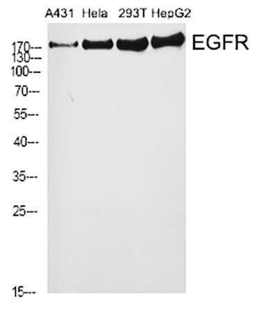
Fig.1. Western Blot analysis of A431 (1), Hela (2), 293T (3), HepG2 (4), diluted at 1:2000.

Fig.2. Immunofluorescence analysis of human liver tissue. 1, EGFR Polyclonal Antibody (red) was diluted at 1:200 (4°C, overnight). 2, Cy3 Labeled secondary antibody was diluted at 1:300 (room temperature, 50min). 3, Picture B: DAPI (blue) 10min. Picture A: Target. Picture B: DAPI. Picture C: merge of A+B.

Fig.3. Immunofluorescence analysis of mouse liver tissue. 1, EGFR Polyclonal Antibody (red) was diluted at 1:200 (4°C, overnight). 2, Cy3 Labeled secondary antibody was diluted at 1:300 (room temperature, 50min). 3, Picture B: DAPI (blue) 10min. Picture A: Target. Picture B: DAPI. Picture C: merge of A+B.
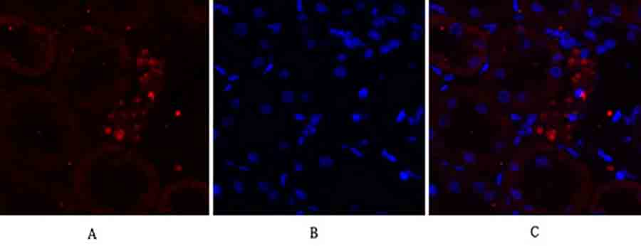
Fig.4. Immunofluorescence analysis of rat kidney tissue. 1, EGFR Polyclonal Antibody (red) was diluted at 1:200 (4°C, overnight). 2, Cy3 Labeled secondary antibody was diluted at 1:300 (room temperature, 50min). 3, Picture B: DAPI (blue) 10min. Picture A: Target. Picture B: DAPI. Picture C: merge of A+B.
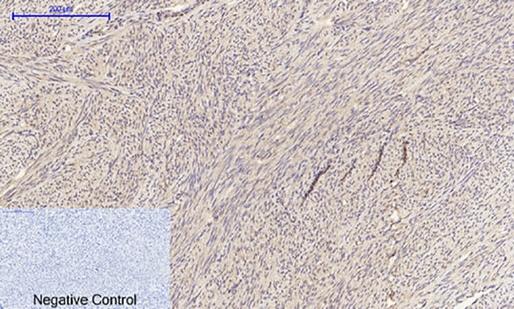
Fig.5. Immunohistochemical analysis of paraffin-embedded human uterus tissue. 1, EGFR Polyclonal Antibody was diluted at 1:200 (4°C, overnight). 2, Sodium citrate pH 6.0 was used for antibody retrieval (>98°C, 20min). 3, secondary antibody was diluted at 1:200 (room temperature, 30min). Negative control was used by secondary antibody only.

Fig.6. Immunohistochemical analysis of paraffin-embedded mouse heart tissue. 1, EGFR Polyclonal Antibody was diluted at 1:200 (4°C, overnight). 2, Sodium citrate pH 6.0 was used for antibody retrieval (>98°C, 20min). 3, secondary antibody was diluted at 1:200 (room temperature, 30min). Negative control was used by secondary antibody only.
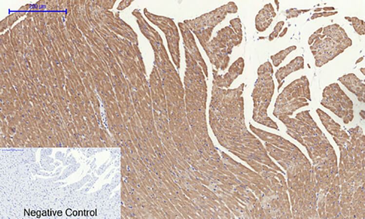
Fig.7. Immunohistochemical analysis of paraffin-embedded rat heart tissue. 1, EGFR Polyclonal Antibody was diluted at 1:200 (4°C, overnight). 2, Sodium citrate pH 6.0 was used for antibody retrieval (>98°C, 20min). 3, secondary antibody was diluted at 1:200 (room temperature, 30min). Negative control was used by secondary antibody only.
You must be logged in to post a review.


Reviews
There are no reviews yet.