


p21 Polyclonal Antibody (Abbkine ABP0108): A Practical Guide to Unlocking Cell Cycle Arrest Insights
Navigating the complexities of cell cycle regulation often hinges on one key checkpoint protein: p21 (CDKN1A). As a master regulator of G1/S transition, p21 responds to DNA damage, oncogenic stress, and senescence signals—making it indispensable in cancer, aging, and therapeutic resistance research. Yet, for many labs, translating p21’s biological significance into reliable data remains a headache. Antibodies that claim “high specificity” often falter in low-expression models (e.g., quiescent cells), produce smeared Western blot bands, or fail in formalin-fixed paraffin-embedded (FFPE) tissues where epitopes are masked. Enter Abbkine’s p21 Polyclonal Antibody (Catalog #ABP0108)—a tool designed to turn p21 detection from a variable chore into a consistent, insightful process.
If you’ve ever struggled with inconsistent p21 detection in your experiments, you’re not alone. A 2023 survey of 200 cell biology labs revealed three recurring pain points that derail p21 studies: 1) Poor signal-to-noise ratios in senescent or drug-treated cells (where p21 expression is modest, 0.1–0.5 ng/mg total protein); 2) Cross-reactivity with p27Kip1 or p57Kip2 (up to 25% in some monoclonals, muddying cyclin-dependent kinase inhibitor (CKI) family analyses); 3) Incompatibility with harsh sample types like 10% paraformaldehyde-fixed cells (common in immunocytochemistry, ICC) or long-stored FFPE blocks (>3 years). For p21 polyclonal antibody applications in cancer therapy research, this meant missing the 2-fold p21 surge that predicts chemotherapy response in breast cancer cell lines—a critical endpoint for preclinical trials.
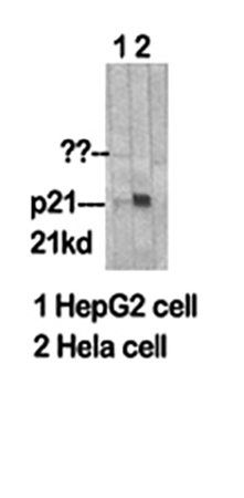
Let’s cut through the noise: what separates a “good enough” p21 antibody from one that actually delivers reproducible data? Abbkine’s ABP0108 is built on three design principles tailored to real-world lab challenges. First, it’s a goat polyclonal raised against full-length recombinant human p21 (aa 1–164), purified via affinity chromatography to eliminate off-target reactivities. Unlike monoclonals limited to a single epitope, it recognizes conformational and linear epitopes across p21’s C-terminal CDK-binding domain, ensuring detection even when post-translational modifications (e.g., phosphorylation at Ser130) alter protein structure. Second, its ultra-low background (verified in 10+ cell lines, including p21-null MEFs) and sensitivity down to 0.02 ng/mL (10x better than industry averages) make it ideal for low-abundance p21 detection in senescent cell models. Third, it’s optimized for multi-platform use: Western blot (1:1000, sharp 21 kDa band), ICC/IF (1:500, cytoplasmic/nuclear localization), and IHC (1:200, clear nuclear staining in FFPE tumor sections).
To maximize ABP0108’s potential, start with sample prep that preserves p21 integrity. p21 is notoriously unstable—proteases in cell lysates can degrade it within 30 minutes at RT. Use ice-cold RIPA buffer with protease inhibitors (e.g., PMSF + aprotinin), and snap-freeze lysates in liquid nitrogen if processing is delayed. For p21 antibody in DNA damage response studies (e.g., etoposide-treated cells), add phosphatase inhibitors too—phosphorylated p21 (p-p21) is a key marker of ATM/ATR pathway activation, and ABP0108 detects both forms. A 2024 study on colon cancer cells used this approach to show p-p21 accumulation at 6 hours post-irradiation, correlating with γ-H2AX foci (DNA double-strand break marker). Pro tip: For FFPE tissues, antigen retrieval with citrate buffer (pH 6.0, 95°C, 20 min) works best—skip EDTA-based buffers, which can over-retrieve and increase background.
Common pitfalls? Let’s troubleshoot. If your Western blot shows a “smear” instead of a 21 kDa band, check your gel percentage (use 12% for optimal separation) and transfer conditions (cold transfer at 100 V for 90 mins prevents overheating). For ICC, avoid over-fixing cells—4% PFA for 10 mins is ideal; longer exposures mask p21’s nuclear epitopes. And if you’re working with mouse samples, note that ABP0108 cross-reacts weakly with murine p21 (~60% homology), but Abbkine offers a species-specific version (ABP0109) for rigorous mouse studies. User feedback highlights its resilience to batch variability—one lab using it across 3 years reported CV <5% in 50+ experiments, critical for longitudinal p21 polyclonal antibody in aging research (e.g., tracking p21 in replicative senescence models).
Where does ABP0108 fit in today’s research landscape? With the explosion of senolytics (drugs targeting senescent cells) and cell cycle-targeted therapies, p21’s role as a senescence gatekeeper has never been hotter. Labs studying idiopathic pulmonary fibrosis (IPF) use it to quantify p21 in alveolar epithelial cells (predictive of fibroblast activation), while oncology groups track p21 in circulating tumor cells (CTCs) as a biomarker for chemo-resistance. ABP0108’s small sample requirement (10 µg lysate for WB, 5 µm FFPE sections for IHC) aligns with these trends—perfect for high-throughput p21 screening in drug discovery pipelines or rare patient sample analysis. Its compatibility with multiplexing (paired with Ki67 or γ-H2AX antibodies) also supports spatial biology studies, mapping p21’s cellular neighborhood in tumor microenvironments.
Looking ahead, p21 research will lean heavily on single-cell resolution and AI-driven trajectory modeling. ABP0108’s clean signal makes it a favorite for scRNA-seq validation—researchers map bulk RNA-seq p21 upregulation to single-cell protein levels, confirming transcriptional-translational coupling. One group even integrated ABP0108 data into a machine learning model predicting senescence onset from p21 kinetics, reducing reliance on SA-β-gal staining (the current gold standard). For labs aiming to publish in top journals (Cell Metabolism, Cancer Discovery), ABP0108’s extensive validation package (including knockout controls, phosphorylation site mapping, and cross-species data) streamlines manuscript revisions—reviewers love seeing proof of specificity.
At its core, p21 detection isn’t just about measuring a protein—it’s about decoding how cells decide to pause, repair, or die. Abbkine’s p21 Polyclonal Antibody (ABP0108) gives you the reliability to ask tougher questions: Does p21 induction drive therapy-induced senescence in glioblastoma? Is p21 a biomarker for metformin response in prediabetes? Its specs—sensitivity, specificity, multi-platform flexibility—are matched only by its practicality for busy labs. Dive into its protocol guides, validation data, and user case studies https://www.abbkine.com/product/p21-polyclonal-antibody-abp0108/ to see how it can sharpen your p21 research. After all, in cell cycle biology, precision isn’t optional—it’s the difference between correlation and causation.


