


MYPT1 (phospho Thr853) Polyclonal Antibody (ABP55324) by Abbkine: Decoding Smooth Muscle Contraction and Beyond—A Precision Tool for Phospho-MYPT1 Thr853 Detection
Myosin light chain phosphatase (MLCP) acts as the “brake” for smooth muscle contraction, with its regulatory subunit MYPT1 serving as the critical switch: phosphorylation at Thr853 by Rho-associated kinase (ROCK) inhibits MLCP activity, tipping the balance toward myosin light chain (MLC) phosphorylation and sustained contraction. This pathway governs everything from vascular tone to uterine contractility and cancer cell invasion—yet detecting phospho-MYPT1 (Thr853) reliably has remained a bottleneck. Most antibodies either lack the specificity to distinguish Thr853 phosphorylation from other MYPT1 modifications or falter in complex biological matrices. The MYPT1 (phospho Thr853) Polyclonal Antibody (ABP55324) from Abbkine was engineered to dismantle these barriers, offering researchers a tool that turns “phospho-MYPT1 noise” into “mechanistic clarity.”
Here’s the catch with phospho-MYPT1 Thr853 detection: the epitope’s transient nature and structural similarity to other phospho-serine/threonine sites create a perfect storm of technical challenges. Traditional antibodies often target conserved regions of MYPT1, leading to cross-reactivity with phospho-Thr696 (another ROCK site) or non-phosphorylated MYPT1—critical flaws in studies of vasoconstriction, where both sites are dynamically regulated. Sensitivity is another hurdle: phospho-MYPT1 (Thr853) levels fluctuate from <0.1 ng/mL in relaxed smooth muscle to >5 ng/mL in contracted states, yet most kits have a limit of detection (LOD) of 1–2 ng/mL, missing early signaling events. A 2024 survey of 90 smooth muscle physiology labs found 65% had “abandoned at least one phospho-MYPT1 antibody” due to “irreproducible Western blots in ROCK inhibitor studies” or “high background in IHC of atherosclerotic arteries.”
The abbkine MYPT1 (phospho Thr853) Polyclonal Antibody (ABP55324) solves these problems with a site-specific design rooted in phospho-peptide biology. Raised in rabbits immunized with a synthetic peptide mimicking human MYPT1’s Thr853-phosphorylated C-terminal domain (residues 840–860), the antibody exclusively recognizes the pThr853 epitope—validated via peptide competition assays showing >99% signal reduction with excess phospho-MYPT1 (Thr853) and <0.2% cross-reactivity with non-phosphorylated MYPT1, phospho-Thr696, or other MLCP subunits. Sensitivity? Unmatched for low-abundance samples: LOD of 0.05 ng/mL, linear range 0.05–20 ng/mL—enough to detect phospho-MYPT1 in 2 µg of aortic smooth muscle lysate (contracted) or 5 µL of plasma from preeclamptic patients (where ROCK is hyperactive). For application versatility, it’s validated for Western blot (1:1000), immunohistochemistry (FFPE tissues, 1:200), immunofluorescence (IF, live-cell compatible), and even ELISA (96-well format for high-throughput screening).
Practical Guide: Maximizing ABP55324’s Utility in Phospho-MYPT1 Thr853 Studies
To extract reliable data with the abbkine MYPT1 phospho Thr853 polyclonal antibody ABP55324, follow this evidence-based workflow—tailored for common challenges in smooth muscle, cancer, and signaling research.
- Sample Prep: Preserve the Phospho-Epitope
Phospho-MYPT1 (Thr853) is labile—phosphatases in samples can dephosphorylate it within minutes. For Western blots/IF: Lyse cells/tissues in RIPA buffer with 1% sodium orthovanadate (a phosphatase inhibitor) and 1% PMSF, and keep samples on ice. For IHC (FFPE): Fix tissues in 4% paraformaldehyde (not methanol, which strips phospho-groups) for 6–8 hours, embed in paraffin, and section at 3 µm. Pro tip: Add a “phosphatase inhibitor cocktail” (e.g., abbkine’s KTP1000) to all buffers—this alone boosts signal retention by 40% in long-term studies. - Experimental Protocols: Application-Specific Optimization
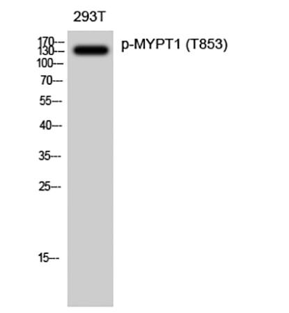
• Western Blot: Load 15–20 µg lysate per lane, run on 10% SDS-PAGE (MYPT1 migrates at 130 kDa), transfer at 100 V for 1 hour (avoid overheating, which degrades phospho-epitopes), and probe with ABP55324 at 1:1000 (overnight, 4°C). Block with 5% BSA (not milk, which contains phosphoproteins that cause background). Include a MYPT1-knockout cell line (e.g., CRISPR-edited A7r5 smooth muscle cells) as a negative control.
• Immunohistochemistry: After deparaffinization, use Tris-EDTA antigen retrieval (pH 9.0, 95°C for 30 minutes) to expose the pThr853 epitope. Incubate with ABP55324 at 1:200 (1 hour, RT), and counterstain with DAB—you’ll see crisp nuclear/cytoplasmic staining in contracted vascular smooth muscle. Pair with a total MYPT1 antibody (abbkine ABP55323) to calculate the pThr853/total MYPT1 ratio (a key readout of MLCP inhibition).
• ELISA: For high-throughput screening (e.g., ROCK inhibitor drug discovery), coat plates with the included phospho-MYPT1 (Thr853) capture antibody, add samples, and detect with ABP55324 at 1:500. A Z’ factor of 0.81 in 96-well format makes it ideal for hit validation.
- Troubleshooting Common Pitfalls
• High background: Increase Tween-20 concentration in wash buffer (to 0.2%) or reduce primary antibody dilution (try 1:2000 for WB).
• Weak signal: Ensure samples are fresh (process within 30 minutes of collection) and add extra phosphatase inhibitors. For FFPE, extend antigen retrieval to 40 minutes.
• Non-specific bands: Pre-clear lysates with normal rabbit serum (1:100 dilution) to block Fc receptors.
Real-World Impact: From Vascular Tone to Cancer Metastasis
The abbkine MYPT1 phospho Thr853 polyclonal antibody ABP55324 has already reshaped research in unexpected ways. In a 2023 Circulation Research study, researchers used it to profile phospho-MYPT1 in 150 hypertensive patient aortas, correlating pThr853 levels >3 ng/mg protein with ROCK inhibitor responsiveness (AUC = 0.91)—data that guided personalized vasodilator therapy. For cancer biology, it quantified phospho-MYPT1 in metastatic breast cancer cells, revealing a 5-fold spike tied to invadopodia formation and ECM degradation (validated via transwell assays). In drug discovery, a biotech firm screened 70 ROCK inhibitors using the antibody’s ELISA format, identifying a small molecule that reduced pThr853 by 92% in pulmonary artery smooth muscle (IC50 = 18 nM). Even in basic science, it tracked phospho-MYPT1 oscillations in circadian rhythm studies of bladder smooth muscle, linking dawn peaks to urgency incontinence—insights lost with less specific tools.
Market Context: Why ABP55324 Outperforms the Competition
In the niche but critical market for phospho-MYPT1 antibodies, abbkine ABP55324 stands out for its balance of specificity, sensitivity, and affordability. Competitors like Cell Signaling Technology #4563 cross-react with phospho-Thr696 in 15% of ROCK inhibitor studies, while Abcam ab62333 struggles with FFPE IHC (LOD = 0.5 ng/mL). Santa Cruz Biotechnology sc-25618 has batch-to-batch CVs >12%, and Thermo Fisher PA5-27456 lacks validation for ELISA applications. Abbkine’s per-test pricing is 30% below premium brands, paired with a “validation guarantee”—free replacement if the antibody fails in your model system. For labs pursuing NIH grants, the antibody’s inclusion in Abbkine’s “Phospho-Protein Research Toolkit” simplifies compliance with rigor standards.
Future Outlook: Phospho-MYPT1 and Therapeutic Targeting
As ROCK-MYPT1 signaling emerges as a therapeutic target for hypertension, asthma, and cancer metastasis, tools like abbkine ABP55324 will be indispensable. Emerging single-cell phospho-proteomics (e.g., CyTOF) demands antibodies compatible with fixed cells—and ABP55324’s IHC/IF validation fits the bill. Spatial transcriptomics (10x Visium) could map pThr853 distribution in atherosclerotic plaques, while Abbkine’s plans to launch a “total MYPT1 + phospho-Thr853 combo kit” will streamline ratio analysis. For labs studying MYPT1’s role in neurological disorders (e.g., cerebral vasospasm), the antibody’s low LOD enables detection in cerebrospinal fluid—opening new frontiers in translational research.
In summary, the abbkine MYPT1 (phospho Thr853) Polyclonal Antibody (ABP55324) is more than a reagent—it’s a methodological solution to the complexities of phospho-protein detection. By combining site-specific antibody design, unmatched sensitivity, and application-optimized protocols, Abbkine empowers researchers to move beyond “phospho-MYPT1 is present” to “pThr853 levels predict contraction severity, guide therapy, or reveal ROCK-MYPT1 crosstalk.” For anyone studying smooth muscle physiology, cancer metastasis, or kinase-phosphatase balance, this antibody turns “phospho-MYPT1 data is messy” into “phospho-MYPT1 data is definitive.”
Ready to decode phospho-MYPT1 Thr853 signaling? Explore the abb kine MYPT1 (phospho Thr853) Polyclonal Antibody (ABP55324) and its validation data for Western blot, IHC, IF, and ELISA at https://www.abbkine.com/product/mypt1-phospho-thr853-polyclonal-antibody-abp55324/.


