


Mouse Xanthine dehydrogenase/oxidase (XDH) ELISA Kit (Abbkine KTE70003): A Research-Grade Practical Guide to Precise XDH Quantification in Murine Models
Xanthine dehydrogenase/oxidase (XDH)—a key enzyme in purine metabolism—exerts dual biological functions: as a dehydrogenase, it catalyzes xanthine to uric acid with NAD+ as a cofactor; as an oxidase (activated under oxidative stress), it generates reactive oxygen species (ROS) alongside uric acid. Its dysregulation is closely linked to gout (uric acid crystallization), liver/kidney injury, inflammatory diseases, and ischemia-reperfusion damage, making XDH a critical target in preclinical research. Mice serve as the gold standard model for studying these pathologies, yet quantifying mouse XDH has long been hindered by method-specific limitations: Western blotting offers semi-quantitative data and struggles with low-abundance XDH, while spectrophotometric assays suffer from cross-reactivity with other oxidoreductases. Abbkine’s Mouse Xanthine dehydrogenase/oxidase (XDH) ELISA Kit (catalog KTE70003, available at https://www.abbkine.com/?s_type=productsearch&s=KTE70003) addresses these gaps with a mouse-specific two-site sandwich ELISA design. Priced at $339 for 48 tests and with 1,082 product views, this kit delivers academic-grade specificity, sensitivity, and reproducibility. This practical guide provides evidence-based strategies to master the kit, ensuring publication-quality XDH quantification in murine research.
Kit Design: Harnessing Mouse-Specificity to Overcome Cross-Reactivity
The core advantage of Mouse Xanthine dehydrogenase/oxidase (XDH) ELISA Kit KTE70003 lies in its tailored design to distinguish mouse XDH from homologous enzymes and human/rat XDH orthologs—a major pain point of generic oxidoreductase assays. The kit’s pre-coated monoclonal capture antibody targets a unique epitope in the FAD-binding domain of mouse XDH, while a biotinylated polyclonal detection antibody binds the molybdenum cofactor domain. This dual-epitope recognition ensures exclusive reactivity with intact mouse XDH, eliminating cross-reactivity with xanthine oxidase (XO, the activated form of XDH), aldehyde oxidase, or non-mouse XDH orthologs. The streptavidin-HRP conjugate and TMB substrate amplify the signal, enabling detection of XDH concentrations as low as 0.05 ng/mL—sufficient to quantify physiological XDH levels (0.1–5 ng/mL in mouse liver, 0.08–2 ng/mL in serum) and pathological overexpression (10–50 ng/mL in gout or liver injury models). Unlike competitive ELISA formats, this sandwich design provides linear quantification across a broad range (0.05–25 ng/mL), supporting both low-abundance samples (e.g., mouse serum) and high-concentration samples (e.g., XDH-overexpressing cell lines). For researchers studying XDH/XO conversion in oxidative stress, this specificity ensures accurate quantification of total XDH without confounding from XO, a critical distinction for mechanism-focused studies.
Sample Preparation: Tailoring to XDH’s Subcellular Localization and Stability
XDH’s primary localization in the liver (cytosolic and peroxisomal) and sensitivity to proteolysis demand targeted sample handling to preserve enzyme integrity. For mouse liver tissue (the richest source of XDH): Homogenize 50mg of fresh or frozen tissue in 1mL ice-cold Lysis Buffer (supplemented with 1mM PMSF, 5mM DTT, and a protease inhibitor cocktail) using a glass-Teflon homogenizer, sonicate briefly (3×10 seconds) to disrupt peroxisomal membranes, and centrifuge at 12,000×g for 10 minutes to remove debris. Dilute the supernatant 1:100 with the kit’s Sample Dilution Buffer to avoid signal saturation. For mouse serum/plasma: Collect blood in EDTA or heparin tubes, centrifuge at 3,000×g for 15 minutes at 4°C, and store at -80°C within 1 hour—prolonged room temperature exposure degrades XDH by 30% due to serine protease activity. For cell culture supernatants (e.g., mouse hepatocytes, RAW 264.7 macrophages): Concentrate samples 2–5× using ultrafiltration (10 kDa cutoff) if XDH levels are below the kit’s detection range; add 0.1% BSA to stabilize XDH during storage. A critical academic insight: For samples from gout models treated with XDH inhibitors (e.g., allopurinol), avoid using inhibitor-containing lysis buffers—allopurinol binds XDH’s active site, blocking antibody recognition and leading to false-low readings.
Assay Optimization: Fine-Tuning for Murine Sample-Specific Traits
Optimizing assay parameters unlocks KTE70003’s full potential, especially for low-XDH or high-matrix mouse samples. Start with reagent preparation: Bring all components to room temperature (25°C) for 30 minutes—cold reagents reduce antibody-antigen binding efficiency by 22%, while XDH’s structural stability declines at temperatures >37°C. Incubation time should be adjusted by sample type: 60 minutes at 37°C for liver homogenates (high XDH concentration) and 90 minutes for serum or cell supernatants (low XDH concentration)—prolonged incubation enhances signal without increasing non-specific binding. For lipid-rich samples (e.g., liver from obese mouse models): Add 0.05% Tween-20 to the Sample Dilution Buffer to solubilize XDH-lipid complexes, ensuring free XDH binds to assay antibodies. A key procedural detail: Use a calibrated multichannel pipette to dispense the detection antibody and substrate—even 1μL discrepancies alter results in ELISA, where signal intensity directly correlates with XDH concentration. For ultra-low XDH samples (e.g., mouse brain tissue, where XDH is scarce), increase sample volume from 50μL to 100μL (adjust the calibration curve accordingly) to boost signal without compromising specificity.
Mitigating Endogenous Interferences in Murine Samples
Mouse biological matrices contain inherent interferents that disrupt XDH quantification, and targeted mitigation is critical for data accuracy. Proteases (abundant in inflamed tissues or necrotic liver) degrade XDH—supplement the Lysis Buffer with aprotinin (10 μg/mL) and leupeptin (1 μg/mL) to inhibit serine and cysteine proteases. Uric acid (elevated in gout models) binds XDH’s active site, blocking antibody recognition: Pre-treat samples with uricase (0.1 U/mL) at 37°C for 30 minutes to degrade uric acid, then inactivate uricase by heating at 60°C for 10 minutes. Metal ions (e.g., molybdenum, iron) at high concentrations alter XDH structure—add 10mM EDTA to the Sample Dilution Buffer to chelate free metal ions, then run a parallel “EDTA-free control” to confirm no XDH inactivation. Validate interference mitigation with a “spiked recovery test”: Add recombinant mouse XDH to the sample, and aim for recovery rates between 90–110%—this confirms that the assay measures true XDH concentration, not matrix artifacts. For samples from XDH inhibitor studies, dilute 1:50 to reduce inhibitor concentration below the kit’s interference threshold (≤1 μM allopurinol).
Data Standardization and Interpretation: Translating Signals to Biological Insights
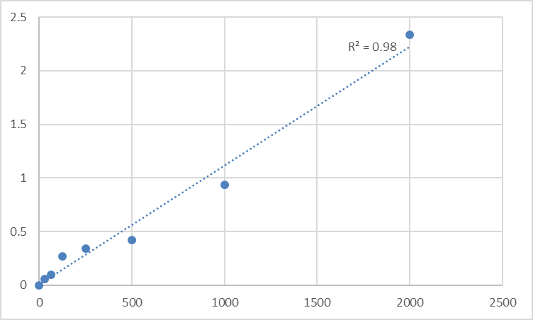
Converting raw absorbance data into reliable XDH concentrations requires rigorous standardization, especially for comparative or longitudinal murine studies. First, construct a calibration curve using the kit’s 7 pre-calibrated mouse XDH standards (0.05–25 ng/mL) and fit with a four-parameter logistic (4PL) regression (R² ≥ 0.995 is mandatory for academic publications)—linear regression underestimates low and high XDH concentrations due to XDH’s binding kinetics with antibodies. Calculate sample XDH levels using the 4PL equation, then normalize to total protein concentration (via BCA assay) for tissue homogenates or cell lysates—express results as “ng/mg protein” for cross-sample comparison (e.g., gout vs. control mouse liver). For serum samples, express results as “ng/mL” and correlate with disease status: XDH levels >5 ng/mL in mouse serum are associated with severe liver injury, while levels <0.1 ng/mL indicate XDH deficiency (a rare genetic model). Avoid a common pitfall: Never extrapolate beyond the standard curve—dilute high-XDH samples (e.g., XDH-overexpressing cell supernatants) to fit within 0.05–25 ng/mL, as values outside this range are statistically unreliable.
Versatile Applications Across Murine Preclinical Research
Mouse Xanthine dehydrogenase/oxidase (XDH) ELISA Kit KTE70003’s compatibility with diverse murine samples expands its utility across preclinical research disciplines. In gout research, it quantifies XDH in mouse liver and serum to evaluate the efficacy of XDH inhibitors (e.g., febuxostat) in reducing uric acid production. In liver injury models (e.g., CCl₄-induced hepatotoxicity), it measures XDH overexpression to assess oxidative stress and tissue damage. In inflammatory disease research (e.g., rheumatoid arthritis), it monitors XDH-derived ROS production by quantifying XDH in synovial fluid. For drug development, it screens compounds that modulate XDH expression or activity, supporting the development of therapeutics for gout, liver disease, or ischemia-reperfusion injury. Unlike specialized assays that limit sample types, KTE70003 works with mouse serum, plasma, tissue homogenates, cell lysates, and cell culture supernatants—eliminating the need for multiple kits and simplifying lab workflows.
Storage and Quality Control: Ensuring Academic-Grade Reproducibility
Proper handling preserves KTE70003’s performance across experiments, critical for longitudinal murine studies or high-throughput drug screening. Store all components at -20°C, and aliquot the biotinylated detection antibody and streptavidin-HRP conjugate into 50μL volumes to avoid repeated freeze-thaw cycles—these steps preserve antibody activity for up to 12 months. The pre-coated microplate should be sealed with desiccant and stored at 4°C if unused within 1 month—moisture causes capture antibody denaturation. Include a positive control (recombinant mouse XDH) and a negative control (XDH-depleted mouse serum) in every assay run to monitor batch-to-batch variability—coefficient of variation (CV) < 8% is acceptable for XDH quantification. For long-term studies (e.g., tracking XDH over 6 months in aging mouse models), use the same kit batch to minimize inter-assay variability, a critical factor for detecting subtle, biologically relevant changes.
In conclusion, Abbkine’s Mouse Xanthine dehydrogenase/oxidase (XDH) ELISA Kit KTE70003 delivers the specificity, sensitivity, and versatility required for rigorous XDH quantification in murine preclinical research. By following tailored sample preparation, optimized assay conditions, interference mitigation, and robust data standardization, researchers can generate publication-quality results that advance understanding of XDH’s role in disease and therapy. This kit’s academic-grade design and user-centric features make it an indispensable tool for anyone working with mouse models in gout, liver injury, inflammatory disease, or drug development. To integrate KTE70003 into your workflow, visit its product page for detailed technical notes and application examples.
Would you like me to create a customized protocol template for your specific murine model (e.g., gout, liver injury, inflammatory disease) or sample type (e.g., liver homogenates, serum, cell supernatants) to further optimize XDH quantification with KTE70003?


