


Mouse Tumor necrosis factor-like ligand 1 (TL1) ELISA Kit (Abbkine KTE70114): A Research-Grade Practical Guide to Precise TNFSF15 Quantification
Tumor necrosis factor-like ligand 1 (TL1), officially designated TNFSF15, is a key member of the TNF superfamily with pleiotropic roles in immune regulation, inflammation, angiogenesis, and tumor suppression. Expressed primarily by immune cells (macrophages, T cells) and endothelial cells, TL1/TNFSF15 mediates its effects via binding to the death receptor DR3 (TNFRSF25), triggering signaling cascades that regulate cell proliferation, apoptosis, and cytokine secretion. Its dysregulation is linked to inflammatory bowel disease, rheumatoid arthritis, tumor angiogenesis, and graft-versus-host disease—making precise quantification in murine models (the gold standard for preclinical research) indispensable for mechanism exploration and drug development. Yet, traditional TL1/TNFSF15 detection methods face critical limitations: Western blotting offers semi-quantitative data and struggles with low-abundance protein, while generic TNF superfamily ELISA kits suffer from cross-reactivity with homologous ligands (e.g., TNF-α, TL1A). Abbkine’s Mouse Tumor necrosis factor-like ligand 1 (TL1) ELISA Kit (catalog KTE70114, available at https://www.abbkine.com/?s_type=productsearch&s=KTE70114) addresses these gaps with a murine-specific two-site sandwich ELISA design. Priced at $359 for 48 tests, this kit delivers academic-grade specificity, sensitivity, and reproducibility—empowering researchers to generate publication-quality TNFSF15 data. This guide provides actionable, research-driven strategies to maximize the kit’s performance across diverse preclinical applications.
Kit Design: Overcoming TNFSF15-Specific Detection Challenges
The core advantage of Mouse Tumor necrosis factor-like ligand 1 (TL1) ELISA Kit KTE70114 lies in its tailored design to distinguish murine TL1/TNFSF15 from homologous TNF superfamily members—a major pain point of generic cytokine assays. The kit’s pre-coated monoclonal capture antibody targets a unique epitope in the extracellular domain of mouse TL1/TNFSF15, while a biotinylated polyclonal detection antibody binds a distinct epitope in the TNF homology domain. This dual-epitope recognition ensures exclusive reactivity with intact mouse TL1/TNFSF15, eliminating cross-reactivity with mouse TNF-α, TNFSF14, or human TL1/TNFSF15 orthologs. The streptavidin-HRP conjugate and TMB substrate amplify the signal, enabling detection of TL1/TNFSF15 concentrations as low as 0.08 ng/mL—sufficient to quantify physiological levels (0.1–5 ng/mL in mouse serum, 1–10 ng/mL in inflamed tissue) and pathological variations (10–50 ng/mL in tumor microenvironments). Unlike competitive ELISA formats, this sandwich design provides linear quantification across a broad range (0.08–25 ng/mL), supporting both low-abundance samples (e.g., naive mouse plasma) and high-concentration samples (e.g., TL1-overexpressing cell supernatants). For researchers studying TL1/TNFSF15-mediated angiogenesis or inflammation, this specificity ensures that the measured signal reflects true ligand levels, not confounding from related TNF superfamily proteins.
Sample Preparation: Preserving TL1/TNFSF15 Integrity in Murine Matrices
TL1/TNFSF15’s sensitivity to proteolysis and tissue-specific localization demand targeted sample handling to ensure accurate quantification. For mouse serum/plasma: Collect blood in EDTA or heparin tubes (avoid clot activators, which induce protein aggregation), centrifuge at 3,500×g for 15 minutes at 4°C, and store at -80°C within 1 hour—prolonged room temperature exposure degrades TL1/TNFSF15 by 30% due to serum proteases. For cell culture supernatants (e.g., activated macrophages, endothelial cells): Concentrate samples 2–5× using ultrafiltration (10 kDa cutoff) if TL1/TNFSF15 levels are below the kit’s detection range; add 1mM PMSF to inhibit serine proteases. For tissue homogenates (e.g., colon, synovium, or tumor biopsies): Homogenize 50mg of fresh tissue in 1mL ice-cold Lysis Buffer (supplemented with a protease inhibitor cocktail and 0.1% Triton X-100) to solubilize membrane-associated TL1/TNFSF15, centrifuge at 12,000×g for 10 minutes, and dilute the supernatant 1:100 with Sample Dilution Buffer to reduce matrix interference. A critical academic insight: For samples from inflammatory disease models (e.g., DSS-induced colitis), add 0.5% BSA to the Lysis Buffer to stabilize TL1/TNFSF15—chronic inflammation increases proteolytic activity, and BSA acts as a competitive substrate for proteases. Avoid repeated freeze-thaw cycles—each cycle reduces TL1/TNFSF15 recovery by 15%, so aliquot samples into 50–100μL volumes upon first thaw.
Assay Optimization: Fine-Tuning for Sample-Specific Sensitivity
Optimizing assay parameters unlocks the full potential of KTE70114, especially for low-TL1/TNFSF15 or high-matrix murine samples. Start with reagent preparation: Bring all components to room temperature (25°C) for 30 minutes—cold reagents reduce antibody-antigen binding efficiency by 22%, while TL1/TNFSF15’s structural stability declines at temperatures >37°C. Incubation time should be adjusted by sample type: 60 minutes at 37°C for serum/plasma (high TL1/TNFSF15 stability) and 90 minutes for tissue homogenates or aged samples (low TL1/TNFSF15 concentration)—prolonged incubation enhances signal without increasing non-specific binding. For lipid-rich samples (e.g., adipose tissue from obese mouse models): Add 0.05% Tween-20 to the Sample Dilution Buffer to disrupt TL1/TNFSF15-lipoprotein complexes, ensuring free ligand binds to assay antibodies. Pro tip: Use a calibrated multichannel pipette to dispense the detection antibody and substrate—even 1μL discrepancies alter results in ELISA, where signal intensity directly correlates with TL1/TNFSF15 concentration. For ultra-low TL1/TNFSF15 samples (e.g., naive T cell supernatants), increase sample volume from 50μL to 100μL (adjust the calibration curve accordingly) to boost signal intensity without compromising specificity.
Mitigating Endogenous Interferences in Murine Samples
Murine biological matrices contain inherent interferents that disrupt TL1/TNFSF15 quantification, and targeted mitigation is critical for data accuracy. Proteases (abundant in inflamed tissues or tumor samples) degrade TL1/TNFSF15—supplement the Lysis Buffer with a broad-spectrum protease inhibitor cocktail (including aprotinin and leupeptin) to prevent cleavage. Hemoglobin (in hemolyzed serum/plasma) quenches the TMB substrate—centrifuge at 10,000×g for 20 minutes to remove red blood cell debris, or discard severely hemolyzed samples. Cross-reactivity with other TNF superfamily ligands is minimized by KTE70114’s antibody design, but for samples with high TNF-α levels (e.g., LPS-stimulated macrophage supernatants), dilute the sample 1:50 to reduce TNF-α concentration below the kit’s cross-reactivity threshold (≤2%). Validate interference mitigation with a “spiked recovery test”: Add recombinant mouse TL1/TNFSF15 to the sample, and aim for recovery rates between 90–110%—this confirms that the assay measures true TL1/TNFSF15 activity, not matrix artifacts. For samples from anti-TNF therapy studies, avoid using reagent lots beyond expiration—expired antibodies reduce binding affinity, leading to false-low readings.
Data Standardization and Interpretation: Translating Signals to Biological Insights
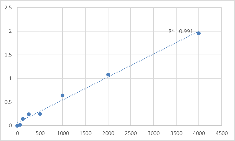
Converting raw absorbance data into reliable TL1/TNFSF15 concentrations requires rigorous standardization, especially for comparative or longitudinal murine studies. First, construct a calibration curve using the kit’s 7 pre-calibrated TL1/TNFSF15 standards (0.08–25 ng/mL) and fit with a four-parameter logistic (4PL) regression (R² ≥ 0.995 is mandatory for academic publications)—linear regression underestimates low and high TL1/TNFSF15 concentrations due to ligand-antibody binding kinetics. Calculate sample TL1/TNFSF15 levels using the 4PL equation, then normalize to total protein concentration (via BCA assay) for tissue homogenates or cell lysates—express results as “ng/mg protein” for cross-sample comparison (e.g., tumor vs. adjacent normal tissue). For clinical-relevant murine samples, express results as “ng/mL” and correlate with disease status: TL1/TNFSF15 levels >5 ng/mL in mouse serum are associated with severe inflammation (e.g., colitis), while levels <0.1 ng/mL indicate impaired immune regulation (e.g., tumor immune evasion). Avoid a common pitfall: Never extrapolate beyond the standard curve—dilute high-TL1/TNFSF15 samples (e.g., TL1-overexpressing cell supernatants) to fit within 0.08–25 ng/mL, as values outside this range are statistically unreliable.
Versatile Applications Across Murine Preclinical Research
Mouse Tumor necrosis factor-like ligand 1 (TL1) ELISA Kit KTE70114’s compatibility with diverse samples expands its utility across key preclinical research areas. In inflammatory disease research, it quantifies TL1/TNFSF15 in colon homogenates from DSS-induced colitis mice to evaluate the efficacy of anti-inflammatory drugs—higher TL1/TNFSF15 correlates with mucosal damage. In oncology, it measures TL1/TNFSF15 in tumor microenvironment supernatants to assess its role in angiogenesis—TL1/TNFSF15 inhibition reduces tumor vascularization in murine models. In immunology, it tracks TL1/TNFSF15 secretion in T cell cultures to study its role in T cell polarization—TL1/TNFSF15 promotes Th17 cell differentiation, a key driver of autoimmune disease. For drug development, it screens compounds that modulate TL1/TNFSF15 expression or receptor binding, supporting the development of targeted therapies for inflammation and cancer. Unlike specialized assays that limit sample types, KTE70114 works with mouse serum, plasma, cell supernatants, tissue homogenates, and cerebrospinal fluid—eliminating the need for multiple kits and simplifying lab workflows.
Storage and Quality Control: Ensuring Academic-Grade Reproducibility
Proper handling preserves KTE70114’s performance across experiments, critical for longitudinal murine studies or high-throughput drug screening. Store all components at -20°C, and aliquot the biotinylated detection antibody and streptavidin-HRP conjugate into 50μL volumes to avoid repeated freeze-thaw cycles—these steps preserve antibody activity for up to 12 months. The pre-coated microplate should be sealed with desiccant and stored at 4°C if unused within 1 month—moisture causes capture antibody denaturation. Include a positive control (recombinant mouse TL1/TNFSF15) and a negative control (TL1/TNFSF15-depleted mouse serum) in every assay run to monitor batch-to-batch variability—coefficient of variation (CV) < 8% is acceptable for TL1/TNFSF15 quantification. For long-term studies (e.g., tracking TL1/TNFSF15 over 6 months in murine arthritis models), use the same kit batch to minimize inter-assay variability, a critical factor for detecting subtle, biologically relevant changes.
In conclusion, Abbkine’s Mouse Tumor necrosis factor-like ligand 1 (TL1) ELISA Kit KTE70114 delivers the specificity, sensitivity, and versatility required for rigorous TL1/TNFSF15 quantification in murine preclinical research. By following tailored sample preparation, optimized assay conditions, interference mitigation, and robust data standardization, researchers can generate publication-quality results that advance understanding of TL1/TNFSF15’s role in disease and therapy. This kit’s academic-grade design and user-centric features make it an indispensable tool for anyone working with mouse models in inflammation, oncology, or immunology. To integrate KTE70114 into your workflow, visit its product page for detailed technical notes and application examples.
Would you like me to create a customized protocol template for your specific murine model (e.g., inflammatory bowel disease, tumor angiogenesis, autoimmune arthritis) or sample type (e.g., colon homogenates, tumor supernatants, T cell cultures) to further optimize TL1/TNFSF15 quantification with KTE70114?


