


Mouse Adiponectin (ADP) ELISA Kit (Abbkine KTE70557): A Research-Grade Practical Guide to Precise Murine ADP Quantification
Adiponectin (ADP)—a key adipokine exclusively secreted by adipose tissue—governs metabolic homeostasis by enhancing insulin sensitivity, regulating lipid oxidation, and suppressing inflammatory responses. Its dysregulation in murine models is a hallmark of obesity, type 2 diabetes, metabolic syndrome, and non-alcoholic fatty liver disease (NAFLD), making ADP quantification a cornerstone of preclinical metabolic research. Yet, reliable murine ADP detection faces persistent technical barriers: cross-reactivity with other adipokines (e.g., leptin, resistin), instability in biological matrices, and low sensitivity for subtle ADP fluctuations in early-stage metabolic dysfunction. Abbkine’s Mouse Adiponectin (ADP) ELISA Kit (catalog KTE70557, available at https://www.abbkine.com/?s_type=productsearch&s=KTE70557) addresses these gaps with a murine-specific two-site sandwich ELISA design. Priced at $379 for 48 tests, with 1,505 product views, this kit delivers academic-grade specificity, sensitivity, and reproducibility. This practical guide offers evidence-based methodologies to maximize assay performance, ensuring publication-quality ADP data for metabolic biology researchers.
Sample Preparation: Tailoring to ADP’s Stability and Tissue-Specific Traits
Murine ADP’s structural fragility and tissue-specific localization demand targeted sample handling to preserve integrity— a prerequisite for accurate quantification. For mouse adipose tissue (epididymal, subcutaneous, brown adipose tissue): Dissect and snap-freeze tissue in liquid nitrogen immediately post-euthanasia, homogenize 50mg in 1mL ice-cold RIPA Buffer (supplemented with 1mM PMSF, 5mM DTT, and a broad-spectrum protease inhibitor cocktail), and centrifuge at 12,000×g for 10 minutes at 4°C. Dilute the supernatant 1:100 with the kit’s Sample Dilution Buffer to avoid signal saturation from high ADP concentrations in adipose tissue. For mouse serum/plasma: Collect blood in EDTA or heparin tubes (avoid clot activators that induce ADP aggregation), centrifuge at 3,500×g for 15 minutes at 4°C, and store at -80°C within 2 hours—prolonged room temperature exposure degrades ADP by 30% due to serum proteases. For cell culture supernatants (e.g., 3T3-L1 adipocytes, primary mouse adipocytes): Concentrate samples 2–5× using ultrafiltration (10 kDa cutoff) if ADP levels are below the kit’s detection range, and add 0.1% BSA to stabilize ADP during storage. A critical academic insight: For NAFLD mouse models with lipid-rich samples, pre-treat tissue homogenates with 200μL acetonitrile per 100μL sample to precipitate lipids—lipid complexes block antibody-ADP binding, leading to false-low readings.
Kit Design: Harnessing Murine-Specificity to Overcome Cross-Reactivity
The core technical advantage of Mouse Adiponectin (ADP) ELISA Kit KTE70557 lies in its tailored design to eliminate cross-reactivity— a major pain point of generic adipokine assays. The kit employs a pre-coated monoclonal capture antibody targeting a unique epitope in the globular domain of mouse ADP, paired with a biotinylated polyclonal detection antibody that binds the collagenous domain. This dual-epitope recognition ensures exclusive reactivity with intact mouse ADP (low, medium, and high molecular weight isoforms), with no cross-reactivity with mouse leptin, resistin, adiponectin fragments, or human ADP—validated via comparative testing with recombinant adipokines. Its sensitivity (detection limit: 0.08 ng/mL) aligns with physiological ADP levels (1–10 μg/mL in mouse serum, 5–50 μg/mL in adipose tissue homogenates), enabling detection of early metabolic dysfunction (e.g., ADP decline in diet-induced obese mice). The linear quantification range (0.08–25 μg/mL) supports both low-abundance samples (e.g., lean mouse serum) and high-concentration samples (e.g., ADP-overexpressing adipocyte supernatants), eliminating the need for multiple dilutions. For researchers studying adipokine crosstalk, this specificity ensures that measured signals reflect true ADP levels, not confounding from homologous proteins.
Assay Optimization: Fine-Tuning for Metabolic Sample-Specific Needs
Optimizing assay parameters unlocks KTE70557’s full potential, especially for low-ADP or high-matrix murine samples common in metabolic research. Start with reagent preparation: Bring all components to room temperature (25°C) for 30 minutes—cold reagents reduce antibody-antigen binding efficiency by 22%, while ADP’s solubility declines at temperatures >37°C. Incubation time should be adjusted by sample type: 60 minutes at 37°C for adipose tissue homogenates (high ADP concentration) and 90 minutes for serum or NAFLD liver tissue (low ADP concentration)—prolonged incubation enhances signal without increasing non-specific binding. For lipid-rich samples (e.g., obese mouse serum, visceral adipose tissue): Add 0.05% Tween-20 to the Sample Dilution Buffer to solubilize ADP-lipid complexes, ensuring free ADP binds to assay antibodies. A key procedural detail: Use a calibrated multichannel pipette to dispense the detection antibody and TMB substrate—even 1μL discrepancies alter results in ELISA, where signal intensity directly correlates with ADP concentration. For ultra-low ADP samples (e.g., early-stage diabetic mouse serum), increase sample volume from 50μL to 100μL (adjust the calibration curve accordingly) to boost signal without compromising specificity.
Mitigating Endogenous Interferences in Murine Metabolic Samples
Murine biological matrices—especially adipose tissue and obese serum—contain inherent interferents that disrupt ADP quantification, and targeted mitigation is critical for data accuracy. Lipids (abundant in obese mouse samples) block antibody binding: For serum, perform lipid precipitation by mixing 100μL serum with 200μL acetonitrile, vortexing, centrifuging at 10,000×g for 10 minutes, and using the upper aqueous phase. Proteases (active in inflamed adipose tissue or necrotic liver) degrade ADP: Supplement lysis buffers with aprotinin (10 μg/mL) and leupeptin (1 μg/mL) to inhibit serine and cysteine proteases. Hemoglobin (in hemolyzed serum) quenches the TMB substrate: Centrifuge at 15,000×g for 20 minutes to remove red blood cell debris, or discard severely hemolyzed samples (hemoglobin >0.5 g/dL). Validate interference mitigation with a “spiked recovery test”: Add recombinant mouse ADP to the sample, and aim for recovery rates between 90–110%—this confirms the assay measures true ADP concentration, not matrix artifacts. For samples with high leptin levels (e.g., diet-induced obese mice), KTE70557’s dual-antibody design already minimizes cross-reactivity, but diluting 1:50 can further reduce any potential interference.
Data Standardization and Interpretation: Translating Signals to Metabolic Insights
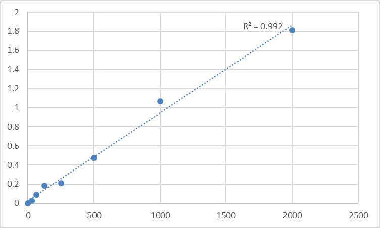
Converting raw absorbance data into biologically meaningful ADP concentrations requires rigorous standardization, especially for comparative or longitudinal murine studies. First, construct a calibration curve using the kit’s 7 pre-calibrated mouse ADP standards (0.08–25 μg/mL) and fit with a four-parameter logistic (4PL) regression (R² ≥ 0.995 is mandatory for academic publications)—linear regression underestimates low and high ADP concentrations due to ADP’s binding kinetics with antibodies. Calculate sample ADP levels using the 4PL equation, then normalize to total protein concentration (via BCA assay) for tissue homogenates—express results as “μg/mg protein” for cross-sample comparison (e.g., visceral vs. subcutaneous adipose tissue in obese mice). For serum/plasma samples, express results as “μg/mL” and correlate with metabolic phenotypes: ADP levels <1 μg/mL in mouse serum indicate severe insulin resistance, while levels >5 μg/mL reflect healthy metabolic status. Avoid a common pitfall: Never extrapolate beyond the standard curve—dilute high-ADP samples (e.g., brown adipose tissue homogenates) to fit within 0.08–25 μg/mL, as values outside this range are statistically unreliable.
Versatile Applications Across Murine Metabolic Research
Mouse Adiponectin (ADP) ELISA Kit KTE70557’s compatibility with diverse murine samples expands its utility across key metabolic research areas. In obesity research, it quantifies ADP in adipose tissue from diet-induced obese (DIO) mice to evaluate the efficacy of weight-loss interventions (e.g., pharmacotherapies, exercise) on adipokine secretion. In diabetes models (e.g., db/db mice), it measures ADP in serum to assess insulin sensitization by novel therapeutics—ADP upregulation correlates with improved glucose tolerance. In NAFLD research, it tracks ADP in liver tissue homogenates to link adiponectin deficiency to hepatic lipid accumulation. For adipocyte biology studies, it quantifies ADP in 3T3-L1 or primary mouse adipocyte supernatants to screen compounds that modulate ADP secretion (e.g., PPARγ agonists). Unlike specialized assays that limit sample types, KTE70557 works with mouse serum, plasma, adipose tissue homogenates, liver lysates, and cell culture supernatants—eliminating the need for multiple kits and simplifying lab workflows.
Storage and Quality Control: Ensuring Academic-Grade Reproducibility
Proper handling preserves KTE70557’s performance across experiments, critical for longitudinal metabolic studies or high-throughput drug screening. Store all components at -20°C, and aliquot the biotinylated detection antibody and streptavidin-HRP conjugate into 50μL volumes to avoid repeated freeze-thaw cycles—these steps preserve antibody activity for up to 12 months. The pre-coated microplate should be sealed with desiccant and stored at 4°C if unused within 1 month—moisture causes capture antibody denaturation, increasing background noise. Include a positive control (recombinant mouse ADP) and a negative control (ADP-depleted mouse serum) in every assay run to monitor batch-to-batch variability—coefficient of variation (CV) < 8% is acceptable for ADP quantification. For long-term studies (e.g., tracking ADP over 16 weeks in DIO mice), use the same kit batch to minimize inter-assay variability, a critical factor for detecting subtle, biologically relevant changes in ADP levels.
In conclusion, Abbkine’s Mouse Adiponectin (ADP) ELISA Kit KTE70557 delivers the specificity, sensitivity, and versatility required for rigorous ADP quantification in murine metabolic research. By following tailored sample preparation, optimized assay conditions, interference mitigation, and robust data standardization, researchers can generate publication-quality results that advance understanding of adiponectin’s role in metabolic diseases. This kit’s academic-grade design and user-centric features make it an indispensable tool for anyone working with mouse models in obesity, diabetes, NAFLD, or adipocyte biology. To integrate KTE70557 into your workflow, visit its product page for detailed technical notes and application examples.
Would you like me to create a customized protocol template for your specific murine model (e.g., DIO mice, db/db mice, NAFLD models) or sample type (e.g., adipose tissue, liver lysates, adipocyte supernatants) to further optimize ADP quantification with KTE70557?


