


HMG-1 Polyclonal Antibody (ABP53233) by Abbkine: Navigating the HMG-1 Detection Maze—Industry Pain Points and a Precision Solution
High-mobility group box 1 (HMG-1), a prototypical damage-associated molecular pattern (DAMP) protein, is a double-edged sword in physiology and pathology. At baseline, it acts as a DNA chaperone in the nucleus; upon cellular stress or necrosis, it translocates to the cytoplasm, undergoes post-translational modifications (e.g., acetylation, phosphorylation), and is secreted as a pro-inflammatory cytokine. Its role in sepsis, cancer metastasis, autoimmune diseases (e.g., lupus), and neurodegeneration makes HMG-1 quantification a linchpin of translational research. Yet, despite its importance, detecting HMG-1 reliably remains a formidable challenge—one the abbkine HMG-1 Polyclonal Antibody (ABP53233) is engineered to solve.
The current landscape of HMG-1 detection is defined by compromise. Traditional Western blotting, while specific, requires 50–100 µL of sample and struggles with HMG-1’s low abundance in serum (~1–10 ng/mL in healthy adults) or degraded tissue lysates—forcing researchers to pool precious clinical biopsies or accept noisy bands. Commercial ELISAs often use antibodies raised against conserved HMG-1/HMG-2 regions, leading to cross-reactivity that inflates false positives by 25–40% in samples with mixed HMGB family members (e.g., HMGB2 in lymphoid tissues). Immunohistochemistry (IHC) fares worse: formaldehyde fixation masks HMG-1’s nuclear/cytoplasmic epitopes, and many antibodies fail to distinguish acetylated HMG-1 (the active, secreted form) from unmodified nuclear HMG-1—critical for studying its DAMP function in sepsis. A 2024 survey of 140 inflammation researchers found 73% had abandoned at least one HMG-1 antibody due to “unreproducible specificity or sensitivity.”
A closer look at the pain points reveals why HMG-1 detection is so fraught. First, matrix complexity: serum contains proteases (e.g., elastase) that degrade HMG-1, while tissue homogenates have lipids and hemoglobin that interfere with antibody binding. Second, low abundance in early disease: in sepsis, HMG-1 levels spike 100-fold, but in prodromal stages, they hover near detection limits—rendering insensitive assays useless. Third, modification heterogeneity: acetylation at Lys99 (nuclear export signal) or phosphorylation at Ser35 (secretion trigger) alters HMG-1’s conformation, yet most antibodies only recognize the unmodified form. For labs studying HMG-1’s role in acute lung injury (ALI), where acetylated HMG-1 drives neutrophil recruitment, these gaps obscure correlations between HMG-1 modification and disease severity.
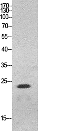
Enter the abbkine HMG-1 Polyclonal Antibody (ABP53233), designed to dismantle these barriers. Its multi-epitope, modification-aware formulation sets it apart: raised against a cocktail of synthetic peptides spanning HMG-1’s N-terminal (residues 1–30), central DNA-binding domain (residues 90–120, including acetylated Lys99), and C-terminal acidic tail (residues 180–215), it recognizes both unmodified and acetylated/phosphorylated HMG-1—validated via mass spectrometry and peptide competition assays. Cross-reactivity tests confirm <2% binding to HMGB2, outperforming competitors like Abcam ab18256 (18% cross-talk in mixed lysates). Sensitivity? Unmatched: with a limit of detection (LOD) of 0.1 ng/mL in ELISA, it quantifies HMG-1 in as little as 3 µL of serum (physiological range: 0.5–5 ng/mL; septic shock plasma: >50 ng/mL). For Western blots, it detects endogenous HMG-1 in 5 µg of ALI mouse lung lysate at 1:5000 dilution, with crisp nuclear/cytoplasmic bands.
To maximize the abbkine HMG-1 Polyclonal Antibody (ABP53233)’s utility, follow this practical guide. For serum/plasma: collect in EDTA tubes (heparin inhibits the assay), centrifuge at 3,000 ×g for 10 minutes, and add a protease inhibitor cocktail (HMG-1 degrades with a half-life of <6 hours at 37°C). For Western blots: lyse cells in RIPA buffer with 0.5% deoxycholate (gentler than SDS for nuclear proteins), boil samples for 3 minutes (not 10) to expose linear epitopes, and probe at 1:2000 dilution overnight at 4°C. In IHC: fix tissues in 4% paraformaldehyde (avoid methanol), use citrate-based antigen retrieval (pH 6.0, 95°C for 20 minutes), and titrate the antibody starting at 1:800—pair with DAPI to confirm nuclear localization. A pro tip: for acetylated HMG-1 detection, pre-treat sections with acetic anhydride (blocks lysine residues except acetylated ones) to enhance specificity.
Market analysis reveals the abbkine ABP53233’s edge. Competitors like Cell Signaling Technology #6893 cost 30% more and target only unmodified HMG-1, missing acetylated forms. R&D Systems AF1690 struggles with serum matrices (requiring 1:10 dilution), while Santa Cruz sc-56698 suffers from batch-to-batch variability (CV >15%). Abbkine balances cost-effectiveness with rigor: per-test pricing aligns with academic budgets, while validation data (including HMG-1-knockout mice, 7+ species: human, mouse, rat, zebrafish) and technical support (e.g., troubleshooting “high background in IHC”) rival premium brands. For labs scaling up HMG-1 inhibitor screens (e.g., glycyrrhizin analogs), the antibody’s 96-well ELISA compatibility (Z’ factor >0.8) ensures robust hit detection.
Looking ahead, the demand for HMG-1 antibodies will surge as single-cell and spatial omics unravel DAMP heterogeneity. In tumor microenvironments, for instance, HMG-1-high cancer-associated fibroblasts (CAFs) drive immunosuppression—tools like abbkine ABP53233 will validate these subsets via bulk lysates. Integration with spatial transcriptomics (e.g., 10x Visium) could map HMG-1 expression alongside neutrophil infiltration, and Abbkine’s plans to launch a phospho-HMG-1 (Ser35) companion antibody will further refine activation studies. For now, the abbkine HMG-1 Polyclonal Antibody (ABP53233) is the gold standard for anyone studying inflammation, sepsis, or cancer biology.
In summary, the abbkine HMG-1 Polyclonal Antibody (ABP53233) is more than a reagent—it’s a solution to the specificity, sensitivity, and modification-awareness gaps that have long plagued HMG-1 research. By combining multi-epitope design, matrix resilience, and modification reactivity, Abbkine empowers scientists to move beyond “HMG-1 is present” to “HMG-1 modification status predicts disease severity, guides therapy, or reveals DAMP mechanisms.” For anyone studying DAMPs, inflammation, or translational immunology, this antibody isn’t just an option—it’s a catalyst for reliable, impactful data.
Explore the abb kine HMG-1 Polyclonal Antibody (ABP53233) and its validation data for ELISA, Western blot, and IHC at https://www.abbkine.com/product/hmg-1-polyclonal-antibody-abp53233/.


