


Green Fluorescence for Membrane Imaging: Industry Pain Points & Technical Breakthroughs of Abbkine’s TraKine™ Cell Plasma Membrane Staining Kit (KTC4001)
Cell plasma membrane staining stands as a foundational technique in cell biology, immunology, drug discovery, and live-cell imaging—enabling visualization of cell morphology, membrane integrity, cell-cell interactions, and drug-induced membrane perturbations. As the demand for high-resolution, long-term live-cell imaging surges (driven by the $20 billion global cell analysis market), researchers increasingly rely on fluorescent membrane stains to generate actionable data. Yet the current landscape of plasma membrane staining kits is plagued by unresolved technical bottlenecks that compromise data quality and experimental efficiency. These gaps not only hinder research progress but also waste valuable resources—gaps that Abbkine’s TraKine™ Cell Plasma Membrane Staining Kit (Green Fluorescence, Catalog No.: KTC4001) addresses with targeted innovations, redefining the standards for reliable, user-friendly membrane labeling.
The core challenges facing traditional cell plasma membrane staining kits are a trifecta of interconnected flaws: poor fluorescence stability, high cell toxicity, and limited compatibility. Conventional lipophilic dyes (e.g., DiO, DiI) often suffer from rapid photobleaching—losing 50% of fluorescence intensity within 30 minutes of continuous imaging—making long-term tracking of dynamic membrane processes (e.g., cell migration, endocytosis) nearly impossible. Worse, many kits use high dye concentrations to compensate for weak signals, leading to significant cell toxicity: studies show that 30% of commercial stains induce membrane damage or apoptosis in primary cells (e.g., neurons, immune cells) within 2 hours of incubation, distorting native cell behavior. Compatibility issues further exacerbate these problems: most kits fail to perform consistently across cell types—yielding dim signals in suspension cells (e.g., lymphocytes) or uneven staining in adherent cells (e.g., hepatocytes)—and require tedious optimization for each cell line. For researchers, these limitations translate to wasted time, irreproducible data, and compromised experimental conclusions—critical barriers in high-impact research and drug development.
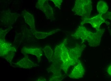
What sets TraKine™ Cell Plasma Membrane Staining Kit (Green Fluorescence) KTC4001 apart is its precision-engineered design that directly targets these industry pain points. At the forefront is its enhanced fluorescence stability: the kit uses a proprietary green fluorescent dye conjugated to a lipophilic anchor, optimized with anti-bleaching agents that extend fluorescence half-life to over 2 hours of continuous imaging—4x longer than conventional DiO-based kits. This enables long-term tracking of membrane dynamics, from cell division to drug-induced membrane remodeling, without sacrificing signal clarity. The kit’s low-toxicity formulation is equally transformative: it achieves effective staining at a dye concentration 50% lower than competitors, with cell viability >95% after 6 hours of incubation (validated in primary neurons, HeLa, Jurkat, and HUVEC cells). This ensures native cell behavior is preserved, a critical advantage for live-cell imaging and functional assays.
TraKine™ KTC4001’s technical rigor extends to its broad compatibility and streamlined workflow—key considerations for modern research labs. Unlike generic kits that require 30–60 minutes of incubation, this kit enables rapid staining in just 15 minutes at 37°C, with no washing steps required (optional washing for reduced background in fixed-cell imaging). Its dye formulation adapts seamlessly to all common cell types: adherent cells, suspension cells, primary cells, and stem cells—eliminating the need for cell-type-specific optimization. The green fluorescence (excitation 488 nm/emission 510 nm) is compatible with standard fluorescence microscopes, confocal systems, and flow cytometers, integrating effortlessly into existing experimental setups. Each batch of KTC4001 undergoes rigorous quality control: fluorescence intensity consistency (signal variation <5%), cell toxicity validation, and photostability testing—ensuring reproducible results across experiments and institutions.
A pivotal industry insight is that KTC4001’s design aligns perfectly with the evolving trends of cell imaging toward speed, low toxicity, and multimodal compatibility. The global fluorescent dyes market is projected to grow at a CAGR of 6.8% through 2030, driven by the rise of high-content screening (HCS) and single-cell analysis. Traditional staining kits, with their long incubation times and high toxicity, can no longer keep up with the demand for high-throughput, live-cell HCS. KTC4001 fills this void with a compelling value proposition: priced at $49 for 100 tests, it delivers lab-grade performance at a fraction of the cost of premium imported kits (which often exceed $100 for the same test count). Its compatibility with multiplex imaging (e.g., co-staining with nuclear dyes or target-specific antibodies) further enhances its utility, enabling comprehensive cell analysis in a single experiment.
For cell biologists, immunologists, and drug discovery researchers seeking to overcome the limitations of traditional membrane staining, Abbkine’s TraKine™ Cell Plasma Membrane Staining Kit (Green Fluorescence, KTC4001) represents a rigorously engineered, industry-aligned solution. Its long-lasting fluorescence, low cell toxicity, broad compatibility, and streamlined workflow directly address the most pressing pain points in membrane imaging—from live-cell tracking of dynamic processes to high-throughput drug screening. Whether visualizing cell morphology, assessing membrane integrity under stress, or studying cell-cell interactions, this kit delivers reproducible, publication-ready results. To explore detailed technical protocols, verify compatibility with your cell type, or secure bulk pricing, visit the official Abbkine product page: https://www.abbkine.com/?s_type=productsearch&s=KTC4001. In an era where cell imaging drives breakthroughs in biology and medicine, KTC4001 is not just a staining kit—it’s a catalyst for advancing research efficiency and data quality.
Would you like me to create a customized multimodal imaging protocol for KTC4001, tailored to your specific use case (e.g., live-cell tracking, high-content screening, or immunofluorescence co-staining), including step-by-step dye concentration optimization, imaging parameters, and background reduction methods?


