| Product name | Anti-α-Tubulin Monoclonal Antibody (3G5) |
| Immunogen | Recombinant Protein |
| Host | Mouse |
| Reactivity | Human, Mouse, Rat |
| Applications | IF, IHC-P, IP, WB |
| Applications notes | Optimal working dilutions should be determined experimentally by the investigator. Suggested starting dilutions are as follows: WB (1:5000-1:10000), IHC-P (1:50-1:300), IF (1:200), IP (1:200). |
| Clonality | Monoclonal |
| Preparation method | The antibody was affinity-purified from mouse ascites by affinity-chromatography using specific immunogen |
| Alternative | TUBA1A; Tubulin alpha-1A chain; Alpha-tubulin 3; Tubulin B-alpha-1; Tubulin alpha-3 chain |
| Formulation | Liquid solution |
| Storage buffer | Liquid in PBS, pH 7.4, containing 0.02% Sodium Azide as preservative and 50% Glycerol. |
| Storage instructions | Stable for one year at -20°C from date of shipment. For maximum recovery of product, centrifuge the original vial after thawing and prior to removing the cap. Aliquot to avoid repeated freezing and thawing. |
| Shipping | Gel pack with blue ice. |
| Precautions | The product listed herein is for research use only and is not intended for use in human or clinical diagnosis. Suggested applications of our products are not recommendations to use our products in violation of any patent or as a license. We cannot be responsible for patent infringements or other violations that may occur with the use of this product. |
| Background | Microtubules of the eukaryotic cytoskeleton perform essential and diverse functions and are composed of a heterodimer of alpha and beta tubulins. The genes encoding these microtubule constituents belong to the tubulin superfamily, which is composed of six distinct families. Genes from the alpha, beta and gamma tubulin families are found in all eukaryotes. The alpha and beta tubulins represent the major components of microtubules, while gamma tubulin plays a critical role in the nucleation of microtubule assembly. There are multiple alpha and beta tubulin genes, which are highly conserved among species. TUBA1A encodes alpha tubulin and is highly similar to the mouse and rat Tuba1 genes. Northern blotting studies have shown that TUBA1A expression is predominantly found in morphologically differentiated neurologic cells. TUBA1A is one of three alpha-tubulin genes in a cluster on chromosome 12q. Mutations in TUBA1A cause lissencephaly type 3 (LIS3) - a neurological condition characterized by microcephaly, mental retardation, and early-onset epilepsy and caused by defective neuronal migration. Alternative splicing results in multiple transcript variants encoding distinct isoforms. |
| Gene ID | 7846/10376 |
| Alternative | TUBA1A; Tubulin alpha-1A chain; Alpha-tubulin 3; Tubulin B-alpha-1; Tubulin alpha-3 chain |
| Accession | Q71U36/P68363 |
| Observed Band(KD) | 52 |
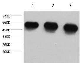
Fig.1. Western blot analysis of 1) Hela, 2) rat brian tissue, 3) mouse brain tissue, diluted at 1:5000.
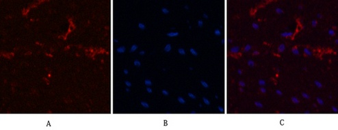
Fig.2. Immunofluorescence analysis of human colon cancer tissue. 1, α-tubulin Monoclonal Antibody (3G5) (red) was diluted at 1:200 (4°C, overnight). 2, Cy3 Labeled secondary antibody was diluted at 1:300 (room temperature, 50min). 3, Picture B: DAPI (blue) 10min. Picture A: Target. Picture B: DAPI. Picture C: merge of A+B.
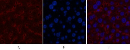
Fig.3. Immunofluorescence analysis of mouse liver tissue. 1, α-tubulin Monoclonal Antibody (3G5) (red) was diluted at 1:200 (4°C, overnight). 2, Cy3 Labeled secondary antibody was diluted at 1:300 (room temperature, 50min). 3, Picture B: DAPI (blue) 10min. Picture A: Target. Picture B: DAPI. Picture C: merge of A+B.
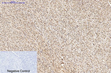
Fig.4. Immunohistochemical analysis of paraffin-embedded human uterus cancer tissue. 1, α-tubulin Monoclonal Antibody (3G5) was diluted at 1:200 (4°C, overnight). 2, Sodium citrate pH 6.0 was used for antibody retrieval (>98°C, 20min). 3, secondary antibody was diluted at 1:200 (room temperature, 30min). Negative control was used by secondary antibody only.
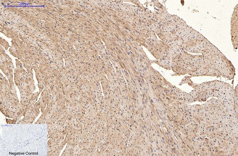
Fig.5. Immunohistochemical analysis of paraffin-embedded mouse heart tissue. 1, α-tubulin Monoclonal Antibody (3G5) was diluted at 1:200 (4°C, overnight). 2, Sodium citrate pH 6.0 was used for antibody retrieval (>98°C, 20min). 3, secondary antibody was diluted at 1:200 (room temperature, 30min). Negative control was used by secondary antibody only.
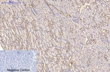
Fig.6. Immunohistochemical analysis of paraffin-embedded rat kidney tissue. 1, α-tubulin Monoclonal Antibody (3G5) was diluted at 1:200 (4°C, overnight). 2, Sodium citrate pH 6.0 was used for antibody retrieval (>98°C, 20min). 3, secondary antibody was diluted at 1:200 (room temperature, 30min). Negative control was used by secondary antibody only.
Author:Li, Jingzhi, et al Publication name:Cell Death & Disease IF:9
Author:Kwon, Yong-Jin, et al. Publication name:mTORC1 pathway IF:5.4
Author:Lee, Hyun-Seung, et al. Publication name:Journal of Ethnopharmacology IF:5.4
Author:Kwon, Yong-Jin, et al. Publication name:Journal of Ethnopharmacology IF:5.4
Author:Kwon YJ, Seo EB, Kim SK, Noh KH, Lee H, Joung YW, Shin HM, Jang YA, Kim YM, Lee JT, Ye SK, Publication name:J Ethnopharmacol IF:3.69
Author:Gao EM, Turathum B, Wang L, Zhang D, Liu YB, Tang RX, Chian RC, Publication name:Reprod Sci IF:2.616
You must be logged in to post a review.


Reviews
There are no reviews yet.